Call Us +65 6884 6788

April 2025


Knee pain when bending is a common issue that can affect people of all ages. Whether it occurs while squatting, climbing stairs, or sitting down, this discomfort can interfere with daily activities. This article explores common causes of knee pain, explaining how different conditions may contribute to discomfort and when medical advice may be needed.

Possible Causes of Knee Pain When Bending

Patellofemoral Pain Syndrome (Runner’s Knee)

Patellofemoral pain syndrome refers to pain around the front of the knee, particularly where the kneecap (patella) meets the thigh bone (femur). It is often linked to repetitive stress or improper kneecap alignment.

Overuse and Repetitive Movements

Activities such as running, cycling, or frequent squatting can place excessive strain on the knee joint, leading to irritation.

Muscle Imbalances

Weak thigh muscles may affect kneecap tracking, increasing stress on the knee. Strengthening exercises for the quadriceps can help stabilise the joint.

Poor Biomechanics

Issues such as flat feet or improper walking posture can contribute to patellofemoral discomfort.

Osteoarthritis

Osteoarthritis occurs when the protective cartilage in the knee joint gradually wears down, leading to pain and stiffness, especially during movement.

Cartilage Degeneration

As the cushioning layer between bones diminishes, friction increases, causing discomfort when bending or straightening the knee.

Morning Stiffness and Swelling

Symptoms may be more noticeable after periods of inactivity and can worsen with prolonged use.

Age and Activity-Related Wear and Tear

Although more common in older adults, osteoarthritis can also develop due to past injuries or prolonged high-impact activities.

Meniscus Tears

The meniscus is a C-shaped soft tissue structure that cushions the knee joint. Tears in this structure can occur due to sudden twisting motions or gradual degeneration.

Acute Injury or Trauma

A meniscus tear often happens during sports or activities that involve sudden knee rotation.

Clicking or Locking Sensation

Some individuals experience difficulty fully extending or bending the knee due to a displaced meniscus fragment.

Pain Along the Joint Line

Discomfort is often felt along the inside or outside of the knee, particularly when squatting or twisting.

Tendonitis (Jumper’s Knee)

Tendonitis occurs when the tendons surrounding the knee become inflamed due to overuse or repetitive strain. The patellar tendon, which connects the kneecap to the shinbone, is most commonly affected.

Pain Just Below the Kneecap

Discomfort often worsens with jumping, running, or prolonged kneeling.

Inflammation and Tenderness

Swelling or sensitivity around the tendon is common, especially after activity.

Gradual Onset

Unlike acute injuries, tendonitis often develops over time due to repetitive stress rather than a sudden event.

Bursitis

Bursae are small fluid-filled sacs that cushion the knee joint. Inflammation of these sacs, known as bursitis, can cause pain and swelling, particularly when bending or kneeling.

Frequent Kneeling or Direct Pressure

Jobs or activities that require prolonged kneeling, such as gardening or carpentry, can increase the risk of bursitis.

Swelling and Warmth Around the Knee

The affected area may appear swollen and feel warm to the touch.

Pain with Movement

Bending the knee or applying pressure can worsen symptoms, making daily tasks uncomfortable.

When to Seek Medical Advice

While occasional knee discomfort may resolve with rest and activity modification, some symptoms indicate the need for medical assessment. Consider consulting a specialist if you experience:

Severe or Persistent Pain

Pain that does not improve with rest or over-the-counter medication may indicate an underlying issue requiring treatment.

“Tightness”, Swelling or Warmth Around the Joint

Significant swelling, redness, or warmth could suggest inflammation, infection, or fluid build-up in the knee.

Clicking, Locking, or Instability

If the knee feels like it might give way, locks in place, or produces a clicking sensation with movement, structural damage may be present.

Difficulty Bearing Weight

If putting weight on the affected leg causes pain or instability, it is best to seek medical attention.

A timely assessment can help determine the cause of knee pain and guide appropriate management.

Conclusion

Knee pain when bending can stem from various conditions, ranging from overuse injuries to degenerative changes. Identifying the cause is the first step in finding effective relief, whether through lifestyle modifications, physiotherapy, or medical treatment.

If knee pain is persistent or interfering with daily activities, schedule a consultation to discuss assessment and management options.

Book Appointment

Fill up the form and we will get back to you soon!

    Synergy Orthopaedic Group

    April 2025

    The anterior cruciate ligament (ACL) plays a key part in knee stability and is often affected by injuries, particularly in sports and activities involving sudden movements. A torn ACL can lead to instability, discomfort, and limitations in daily activities or physical pursuits. In many cases, surgery is considered to improve knee function and support a return to an active lifestyle.

    This guide provides a thorough overview of ACL surgery, covering when it may be considered, the different surgical techniques available, the preparation process, and the recovery journey.

    The ACL and Its Function

    The ACL is one of four primary ligaments in the knee joint, connecting the femur (thigh bone) to the tibia (shin bone). It provides stability by controlling excessive forward movement of the tibia and supporting rotational movements. The ligament is particularly engaged during activities involving sudden stops, directional changes, and jumping.

    When the ACL is torn, knee stability may be affected, resulting in difficulties with movement and an increased risk of further knee damage. Depending on the severity of the injury and lifestyle demands, ACL surgery may be an option to restore knee function.

    When Might ACL Surgery Be Considered?

    Surgical reconstruction of the ACL is typically recommended when non-surgical approaches do not provide sufficient stability. Some common scenarios where surgery may be advised include:

    Knee instability in daily movements

    If the knee frequently gives way during routine activities, ACL surgery may be considered to improve stability. Persistent instability may also contribute to additional knee injuries, such as damage to the meniscus or cartilage.

    Return to sports or active pursuits

    Individuals participating in activities requiring sudden turns, jumps, or rapid changes in movement may require surgical reconstruction to improve knee function and reduce the risk of further injury.

    Recurrent or complex ACL injuries

    Repeated ACL tears or injuries affecting other knee structures may be managed more effectively with surgery compared to non-surgical treatment options.

    In general, there are 2 main reasons for considering ACL surgery:

    • In the short term, it provides for a stable knee to return to an active lifestyle.
    • In the long term, it provides for a stable knee which reduces the risk of a rapid degeneration of the knee over time.

    ACL Surgery Techniques

    ACL surgery involves different approaches and graft options, with the choice depending on the severity of the injury and patient-specific factors.

    ACL Reconstruction vs ACL Repair

    ACL Reconstruction

    This approach replaces the damaged ligament with a graft and is the most widely performed procedure. It is typically recommended for complete or longstanding ACL tears, as it provides a reliable outcome for restoring knee function.

    ACL Repair

    In some cases, the torn ligament can be reattached rather than replaced. This technique is generally considered for recent, partial tears where the remaining ligament tissue is in good condition. However, it is not suitable for all injuries.

    Graft Options for ACL Surgery

    Autografts (patient’s own tissue)

    A graft may be taken from the patellar tendon, hamstring tendon, or quadriceps tendon. These grafts provide strong tissue for reconstruction, though recovery may take longer due to healing at the donor site.

    Allografts (donor tissue)

    Sourced from a donor, allografts eliminate the need for a second surgical site. They may be a suitable option for some individuals, though integration into the knee can take longer.

    Preparing for ACL Surgery

    Proper preparation before ACL surgery can help optimise the recovery process. Key steps include:

    Medical Examination and Assessment

    A physical examination is performed to assess knee stability, range of motion, and any swelling or tenderness. Tests such as the Lachman test or pivot shift test help determine the extent of ligament damage and any associated knee conditions.

    Imaging (MRI and X-ray)

    MRI scans provide detailed images of the ACL and surrounding structures, allowing the surgeon to plan the procedure effectively. X-rays may also be used to assess any involvement of bone structures.

    Pre-Operative Requirements

    Blood tests and other screenings may be conducted to assess overall health before surgery. Medications that affect blood clotting may need to be reviewed, with temporary adjustments made as required. Strengthening exercises may also be recommended before surgery to support recovery.

    What Happens During ACL Surgery?

    The surgical procedure follows a structured sequence of steps to ensure the graft is correctly positioned and secured.

    Cleaning and Preparing the Joint

    The damaged ligament remnants and any loose cartilage or debris are removed to create a clear surgical field. This step helps ensure there are no obstructions to graft placement.

    Arthroscopic Assessment

    A small camera (arthroscope) is inserted into the knee joint through a keyhole incision, providing a detailed view of the affected structures. This allows the surgeon to assess the extent of the injury and address any additional damage, such as meniscus or cartilage injuries.

    Graft Preparation and Placement

    The selected graft is prepared and shaped to match the knee’s dimensions. Small tunnels are created in the femur and tibia, allowing the graft to be positioned in line with the original ACL.

    Securing the Graft

    The graft is fixed in place using screws or other fixation devices to provide stability. The surgeon then tests the graft’s position and tension to ensure it allows for both movement and support.

    Final Inspection and Closure

    After confirming that the graft is secure, the surgeon inspects the joint before closing the incisions with sutures. A bandage is applied to minimise swelling and protect the knee during the initial healing phase.

    Recovering After ACL Surgery

    Recovery from ACL surgery requires a structured rehabilitation programme. Physiotherapy plays a key part in regaining knee function, improving mobility, and reducing the likelihood of complications.

    Early Post-Operative Care

    Crutches and a knee brace are typically used to limit movement and protect the graft during the early healing phase. Pain management may involve prescribed medication and cold therapy to help control discomfort and swelling. Elevating the leg can assist in reducing swelling and preventing stiffness. Follow-up appointments allow the surgeon to monitor progress and adjust care as required.

    Recovery Timeline

    Recovery progresses in phases, with each stage focusing on specific goals. During the first six weeks, the priority is reducing swelling, protecting the graft, and restoring basic movement through gentle exercises. From six to twelve weeks, muscle strengthening exercises are introduced, and weight-bearing increases gradually. By three to six months, advanced physiotherapy, including sports-specific exercises, may be introduced to support a return to full activity. Safe return to full activities, including contact sports, will typically be close to a year post-surgery.

    Potential Risks and Considerations

    ACL surgery is generally well tolerated, but as with any procedure, there are some associated risks. These include infection, bleeding, nerve injury and reactions to anaesthesia, which are managed with appropriate care. In some cases, the graft may not fully integrate, requiring further treatment. Knee stiffness can develop if rehabilitation is inadequate, but this can often be improved through physiotherapy.

    Reducing the Risk of Future ACL Injuries

    Strengthening the muscles around the knee, particularly the quadriceps and hamstrings, can help support the joint. Physiotherapy or structured exercise programmes focusing on strength, flexibility, and movement techniques may reduce the risk of re-injury. Wearing appropriate footwear and avoiding sudden, high-risk movements can also contribute to knee stability.

    Conclusion

    ACL surgery is a recognised method for improving knee stability following ligament injuries. With appropriate preparation, a structured rehabilitation plan, and commitment to recovery, many individuals regain mobility and return to their desired activities.

    If you are considering ACL surgery, schedule a consultation to discuss your treatment options and learn more about the procedure and recovery process.

    Book Appointment

    Fill up the form and we will get back to you soon!

      Synergy Orthopaedic Group

      April 2025

      Running subjects the knee joints to repeated movements that may cause substantial strain over time. Patellofemoral pain syndrome—commonly known as runner’s knee—affects numerous athletes and fitness enthusiasts. This comprehensive guide examines the condition in depth, providing detailed information on its causes, symptoms, diagnosis, treatment options, and preventative techniques to help runners stay active while minimising knee discomfort.

      What is Runner’s Knee?

      Runner’s knee develops when the kneecap (patella) undergoes undue or incorrect pressure while tracking along the groove in the thigh bone (femur). This misalignment generates pain surrounding or behind the kneecap, notably during activities like running, ascending stairs, or performing squats. The condition may emerge progressively or result from overuse or improper biomechanics.

      Recognising Symptoms and Signs

      The symptoms of runner’s knee can vary, but generally include the following:

      Kneecap Pain

      A persistent ache or soreness appears beneath or around the patella, especially during activities that require repeated knee flexion, such as running, squatting or climbing stairs.

      Grinding Sensations

      The knee may produce grinding, clicking, or popping noises when flexing or extending the leg.

      Post-Sitting Discomfort

      Discomfort tends to intensify following prolonged periods of sitting with bent knees, commonly termed the ‘theatre sign’.

      Mild Swelling

      Slight swelling or puffiness may occur around the kneecap, especially after physical exertion.

      Contributing Causes of Runner’s Knee

      Several factors can influence the development of runner’s knee, including:

      Biomechanical Factors

      Flat feet, high arches, knock knees, or other alignment variations may disrupt proper patellar tracking.

      Training Progression Errors

      Sudden increases in running distance, pace, or intensity can overwhelm knee structures, resulting in inflammation or irritation.

      Muscle Imbalances

      Weakness in the hip abductors, gluteal muscles, or quadriceps, coupled with tight hamstrings or calves, can compromise knee alignment and stability.

      Footwear Considerations

      Worn-out or unsuitable running shoes may fail to distribute impact properly, increasing stress on the knee joints.

      Diagnostic Approaches for Runner’s Knee

      An accurate diagnosis of runner’s knee is necessary for effective treatment. A variety of methods are used to assess the condition, including:

      Physical Examination

      A thorough assessment of knee alignment, flexibility, and movement is conducted. Strength and stability tests are performed to identify weaknesses in the surrounding muscles while observing pain patterns during movements such as squatting or stepping.

      Medical History

      An in-depth examination of the patient’s running patterns, past injuries, and symptom progression offers important information about the contributing factors. Enquiries frequently focus on any previous knee problems or recent training modifications.

      Imaging Studies

      X-rays or MRI scans may be recommended to rule out alternative conditions, such as ligament tears or cartilage damage. These images also assist in assessing patellar alignment and overall joint health.

      Effective Treatment Strategies

      Most instances of runner’s knee can be managed effectively through conservative methods.

      Rest and Activity Modification

      Temporarily reducing or modifying high-impact activities allows for knee healing. Low-impact alternatives such as swimming or cycling may be appropriate during recovery. The rest period is tailored based on symptom severity.

      Targeted Strengthening Exercises

      Specific exercises strengthen the quadriceps, hip stabilisers, and core muscles. These programmes focus on enhancing muscular coordination, improving flexibility, and rectifying biomechanical imbalances to decrease stress on the patella.

      Ice Therapy

      Applying a cold compress or ice pack to the knee following activity helps decrease swelling and relieve discomfort. This can be implemented several times daily for 15–20 minutes.

      Pain Management

      Over-the-counter anti-inflammatory medications, such as ibuprofen, may be recommended to address acute pain and inflammation during the initial treatment phase.

      Prevention Strategies for Runners

      Preventing runner’s knee requires adopting practices that reduce knee stress while maintaining proper alignment and muscular balance. The key strategies include:

      Gradual Training Progression

      Build mileage gradually to allow knees and surrounding muscles to adapt properly. A recommended guideline is to increase weekly mileage by no more than 10%.

      Regular Strength Training

      Incorporate exercises targeting the hip, quadriceps, and core muscles into your routine. This promotes better joint alignment and stability, reducing the likelihood of knee issues.

      Proper Footwear Rotation

      Replace running shoes if they display signs of wear. Ensure shoes provide adequate cushioning and arch support suited to your foot type.

      Surface Variation

      Running on different surfaces, such as trails, tracks, and pavement, helps distribute stress more evenly across the joints.

      By prioritising these preventative strategies, runners can maintain their activity levels while lowering their chances of developing knee pain.

      When Medical Attention Is Recommended

      If home remedies do not alleviate symptoms of runner’s knee, professional medical attention may be necessary. Persistent pain that intensifies over time, substantial or increasing swelling, and difficulty walking or bearing weight on the affected leg are all indications to consult a healthcare provider.

      Additionally, symptoms such as the knee unexpectedly giving way or locking during movement may signify underlying structural issues that require further evaluation. Seeking timely medical advice not only ensures an accurate diagnosis but also helps prevent potential complications that could extend recovery or necessitate more intensive treatments.

      Conclusion

      Runner’s knee represents a common but manageable condition affecting athletes and active individuals. By comprehending its causes and symptoms, implementing effective treatment approaches, and practising preventative measures, most people can recover fully and resume their running routines without long-term restrictions. Addressing the issue promptly and consistently is fundamental to ensuring ongoing knee health.

      Schedule a consultation today for a professional assessment and a customised treatment plan to address your running-related knee concerns.

      Book Appointment

      Fill up the form and we will get back to you soon!

        Synergy Orthopaedic Group

        July 2021

        Here’s What You Need To Know

        Few injuries are as frustrating as knee injuries, especially when you’re left sidelined from your favourite sporting activities. Unfortunately, anyone can fall victim to knee injuries regardless of whether they are professional athletes or regular fitness enthusiasts. In fact, knee injuries can also arise from everyday activities or accidents.

        According to a 2006 study, approximately 40% of sports injuries comprise injuries related to the knee joint. But don’t worry, because fixing knees is my specialty. I’ve studied their design, how they function, common injuries, the most cutting edge prevention techniques, and what leading physiotherapists are doing to rehabilitate knee injuries. 

        Regardless of whether your knee injury stems from patellofemoral pain syndrome, ligament sprains or tears, cartilage tears, tendinitis, or just plain old wear and tear (arthritis), this playbook will explain everything to you and offer tips on how you can prevent them.

        If you’ve ever suffered from a knee injury, then this is the article for you to learn more about how you can keep your knees as healthy as possible. 

        If you’ve never had a knee injury before, this article is also for you. By following the injury prevention tips here, your chances of sustaining a knee injury will be greatly lowered.

        Types Of Knee Injuries

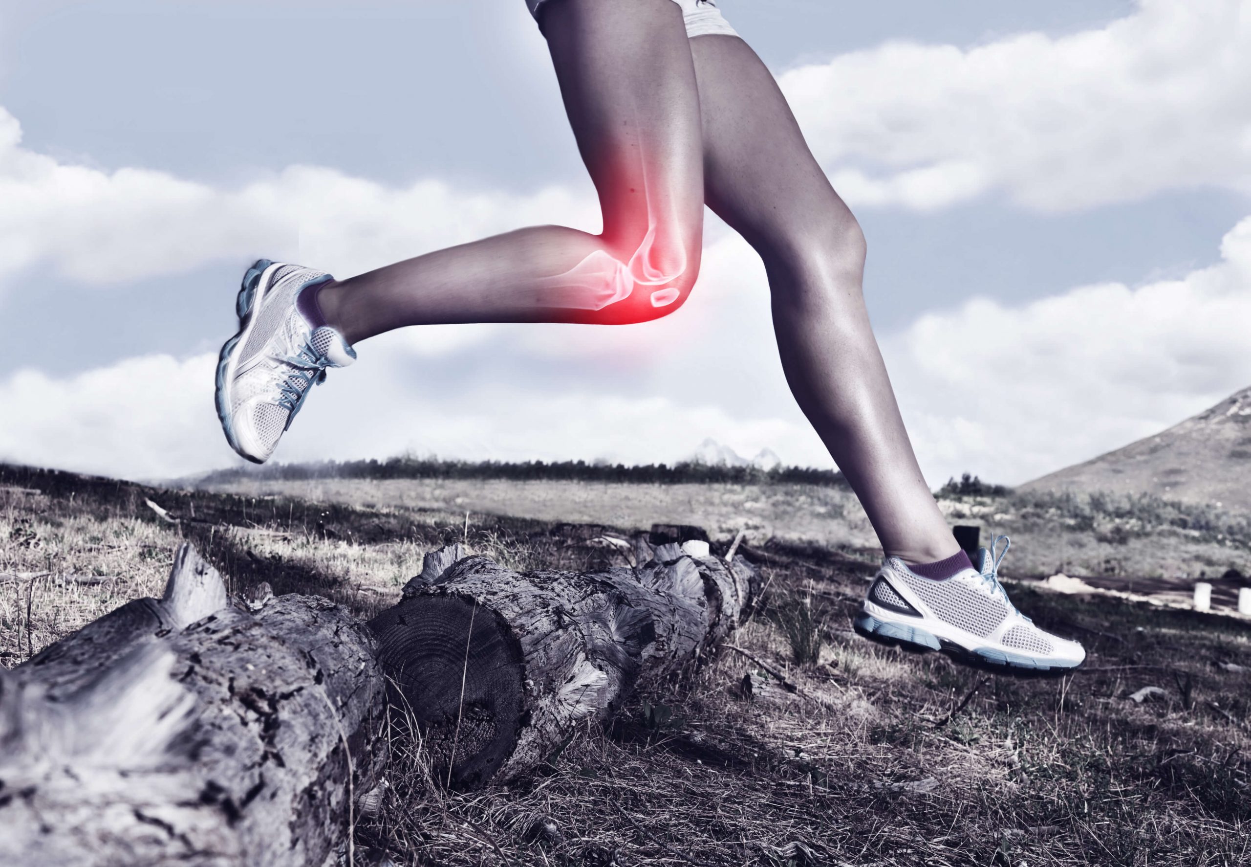
        Before that, you should know a little bit about the anatomy and inner workings of the knee joint in order to understand the different injuries that may occur. The bones at the knee joint consist of the femur (thigh bone), the tibia (shin bone), and the patella (kneecap). The ligaments, which are fibrous connective tissue connecting the bones, help provide joint stabilisation.

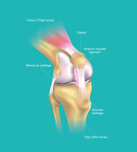
        Here’s a quick guide to help visualise the bones which make up your knee:

        • The femur: runs from your thigh to your knee
        • The tibia: runs from your knee to your ankle
        • The patella: the triangular-shaped bone in front of the knee

        Now let’s take a closer look at the muscles, tendons, and cartilages that make up the knee joint.

        The medial collateral ligament (MCL) provides a form of restraint against valgus forces (anything bent or twisted away from the midline) to the knee. The lateral collateral ligament (LCL), however, provides restraint to varus forces (the opposite of valgus forces) at the knee. These two collateral ligaments are responsible for controlling the sideways motion of your knee and bracing it against unnatural movement.

        Next, the anterior cruciate ligament (ACL) serves as the primary restraint to anterior tibial translation on the femur. The posterior cruciate ligament (PCL), on the other hand, limits posterior tibial translation on the femur. Both of these ligaments cross inside the knee joint, thus earning the name ‘cruciate’ (cross-shaped). More importantly, they help to stabilise the knee from the back and the front.

        There are many different knee injuries. These can be categorised into broken bone injuries (fractures), soft tissue injuries (dislocations, sprains, ACL injuries, PCL injuries, collateral ligament injuries, meniscal tears, and tendon tears), and overuse injuries that build up over time (arthritis).

        ACL Injuries

        An ACL injury is essentially a tear or sprain of the anterior cruciate ligament (ACL), which is the ligament that connects your femur to your tibia.

        Many patients have reported hearing a ‘pop’ sound coming from their knee upon sustaining an ACL injury. This type of injury is most common in intermittent sprint sports which involve sudden changes in direction, such as basketball, hockey, American football, soccer, and rugby amongst others. However, ACL injuries can also occur from accidents such as falling from a height which places sudden pressure on the knee in an awkward position.

        A good number of ACL injuries also come with accompanying damage to other structures in the knee, such as the cartilage, meniscus, and other ligaments. ACL injuries are graded on a severity scale:

        • Grade 1 Sprains: Ligament has been stretched, resulting in mild damage. However, the knee joint is still stable.
        • Grade 2 Sprains: Ligament is stretched until it has become loose. This is commonly referred to as a partial ACL tear.
        • Grade 3 Sprains: Ligament has been split into two pieces, resulting in loss of stability at the knee joint. This is also known as a complete ACL tear.

        Causes

        There are a number of specific movements that can put stress on the knee, resulting in an ACL injury. These include:

        • A sudden stop or change in direction
        • Receiving a blow to the knee during a tackle, resulting in hyperextension or inward bending
        • Pivoting sharply with one foot firmly planted
        • An awkward landing from a jump

        Although sports injuries are the common cause of ACL injuries, other patients have also sustained such injuries during car accidents or other trauma, such as falling down the staircase.

        Symptoms

        Patients who experience an ACL sprain or tear are likely to report some combination of the following symptoms:

        • Sharp and intense pain
        • Rapid swelling
        • Deep aching pain in the knee
        • Knee instability
        • Loss of full range of motion

        Knee Fractures

        A knee fracture is a damage to any of the four leg bones which meet at the knee. This includes the patella, the femur, the tibia, and the fibula. Knee fractures can be as mild as a small hairline crack in the bone, or as severe as a complete break.

        Orthopaedic surgeons categorise fractures in several ways:

        • Non-displaced fracture: Broken pieces of bone remain in contact with each other.
        • Displaced fracture: Bone pieces are out of position and no longer aligned.
        • Comminuted fracture: Bone breaks into many pieces.
        • Open or closed fracture: Denotes whether the bone has broken through the surface of the skin (open) or not (closed).

        Knee fractures are typically caused by direct trauma to the knee from a high impact activity. This may include a collision during a contact sport such as American football, rugby, or judo. Motorcycle accidents may also cause knee fractures when the rider is flung from the motorcycle, resulting in crushing the knee. Weakened bones or diminished bone density can also predispose patients to an increased risk of fractures.

        Causes

        Several causes of knee fractures include:

        • Falling down directly on the knee
        • Receiving a powerful blow to the knee, such as during a car accident
        • Severe and sudden muscle contractions that pull apart the patella

        Symptoms

        Some of the common symptoms of knee fractures include:

        • Extreme pain in the knee
        • Swelling and bruising
        • Tenderness at the knee area
        • Inability to bend or straighten the knee in an extended leg raise
        • Muscle spasms
        • Cracking or popping sound during movement
        • Inability to walk

        Torn Meniscus

        A meniscus tear is another common knee injury reported by patients. Both your knees contain two C-shaped pieces of cartilage which serve as a cushion between your tibia and femur. This is called the meniscus

        To understand how this injury arises, you first need to know what the role of the meniscus is. As mentioned, the menisci sit between the tibia and femur, stabilising the lower part of the leg against forces generated by your bodyweight. The medial meniscus sits on the inside of the knee while the lateral meniscus sits on the outside of the knee.

        Essentially, the meniscus plays an important role in stabilising and distributing load in the knee joint. Meniscus tears occur when a patient twists or turns their upper leg while their foot is firmly planted and their knee is bent awkwardly. This can occur in activities such as dancing, cheerleading, snowboarding, or racquetball.

        You should know that meniscus tears often occur with ACL tears, due to the nature of the knee twisting during the accident. According to a research study carried out by the Shelbourne Knee Centre, approximately 50% of patients with ACL tears also suffer from meniscus tears.

        Causes

        Causes of a torn meniscus include:

        • Forceful twisting or rotating of the knee
        • Sudden pivoting or turning when running
        • Deep squatting or heavy lifting in the gym

        In some cases, degenerative changes to the cartilage in the knee joint can also cause a meniscus sprain or tear with little trauma. This is due to the cartilage slowly losing its blood supply and resilience over time due to natural wear and tear. In other words, the risk of developing a torn meniscus increases with age.

        Symptoms

        The symptoms of a torn meniscus include:

        • Intense pain in the knee joint, usually on the inside, outside, or back of the knee
        • Swelling at the knee area
        • Catching or locking of the knee joint
        • Inability to bend or fully extend the knee
        • Limping while walking

        The pain may be bearable initially but will progressively worsen as inflammation and swelling begin to set in.

        Knee Bursitis

        Knee bursitis is an inflammation of the bursa. Bursas are the thin fluid-filled sacs that sit between your tissue, bones, and knee joint. Other joints such as the hips, shoulders, elbows, and ankles also have bursas which help to reduce friction between the bones and tissues.

        Any one of the bursa in your knee can become inflamed, but knee bursitis most typically occurs over the kneecap or on the inner side of your knee. When these sacs become irritated, it fills with fluid, causing the sac to swell. This is your body’s natural defence mechanism against trauma to the knee by providing extra cushioning and support for the underlying bone.

        Bursitis can occur due to repetitive movements which inflict trauma to the knee. One example of this is kneeling for extended periods of time, especially on a hard surface, resulting in pressure being applied to the knee area. Plumbers, roofers, carpet layers, coal miners, and gardeners have a higher risk of developing this condition.

        However, knee bursitis is not as severe as compared to other knee injuries and usually goes away on its own. This may take up to a few weeks depending on the severity of the injury. The most important factor in the healing process is for you to rest as much as possible by giving the knee a break from whatever was causing the inflammation.

        Causes

        The causes of knee bursitis are:

        • Frequent and sustained pressure on the knee, such as kneeling for an extended period of time
        • Strenuous activity leading to overuse of the knee
        • A direct blow to the knee resulting in mini-trauma
        • Bacterial infection in the bursa
        • Complications arising from osteoarthritis or rheumatoid arthritis of the knee

        Symptoms

        The symptoms of knee bursitis may include:

        • Pain during activity, but no pain at night
        • Swelling and inflammation at the front of the knee
        • Tenderness at the affected area
        • Bursitis caused by bacterial infection may produce pus as well as fever

        Patellar Tendinitis

        Patellar tendinitis, also known as jumper’s knee, is an overuse injury characterised by inflammation of the patellar tendon. Contrary to what its name suggests, the patellar tendon is actually a ligament.

        To understand this injury, we first have to understand the role of the patellar tendon in the knee joint. The patellar tendon connects your patella to your tibia and works with the muscles in the front of your thigh to straighten your leg. Damage to this tendon can make it difficult to move around, hindering you from participating in sports activities.

        Patellar tendinitis occurs when you overuse your knee joint, for example, when you jump and land repeatedly on hard surfaces. It is a type of sports-related injury commonly associated with elite athletes. With repeated stress and impact placed on the knee, your patellar tendon becomes irritated and inflamed from the minor strains and tiny tears. Basketball, netball, and volleyball players are more susceptible to this injury.

        This leads to pain, soreness, and discomfort at the knee area, which may either be minor or severe depending on the extent of damage inflicted. If you continue to exercise or partake in physical activity with this injury, it may worsen over time.

        Causes

        The causes of patellar tendinitis are:

        • Any kind of repeated activity which gradually damages the knee
        • Sports that involve repeated jumping and landing, especially without bracing the knee properly
        • Training on hard surfaces such as concrete

        Physically active athletes who do a lot of running and jumping are more likely to get patellar tendinitis. Other risk factors include having tight quadriceps and hamstring muscles, which can increase the strain placed on your patellar tendon. Lastly, having a muscular imbalance can cause an uneven pull, resulting in tendinitis as well.

        Symptoms

        The symptoms of patellar tendinitis include:

        • Pain between your kneecap and shinbone
        • Tenderness to the touch at the affected area
        • Tightness or stiffness at the knee area, particularly first thing in the morning

        Arthritis

        Many of us think of arthritis simply as an age-related disease that adults become susceptible to in their old age. However, did you know that arthritis as a disease actually comprises a complex family of over 100 types of musculoskeletal disorders?

        Arthritis is a degenerative disease where inflammation and pain affect the joint as well as its surrounding tissues. The most common early signs of arthritis are typically observed in the back, knees, hips, and elbows. Depending on the severity of the condition, the pain and stiffness will vary.

        There are four main types of arthritis:

        • Osteoarthritis: The most common type of arthritis caused by a degradation of the cartilage in your joints over time.
        • Rheumatoid arthritis: An autoimmune disease that can occur at nearly any age.
        • Psoriatic arthritis: Usually observed in patients with psoriasis, an autoimmune skin condition.
        • Gout arthritis: A type of arthritis due to deposits of uric acid crystals in a joint, typically starts in the big toe before moving up the foot and ankle joint.

        In addition, post-traumatic arthritis can arise due to a sports injury or road accident. All of the different types of arthritis above affect the joints in different ways.

        You should know that the damage sustained by the knee joint from osteoarthritis is irreversible. However, the symptoms of pain can be managed by receiving medical treatment, thereby slowing down the progression of the disease.

        Causes

        Osteoarthritis occurs when the cartilage that cushions your bones deteriorate over time. For this reason, osteoarthritis has also been referred to as a ‘wear and tear’ disease that tends to happen with older age. This wear and tear can be hastened by repeated impact injuries or bacterial infection.

        Rheumatoid arthritis, on the other hand, is an autoimmune disease that has no known cause. The body’s immune system attacks the inside lining (synovial membrane) of the joint capsule, resulting in inflammation and swelling. It is thought to be due to hereditary factors that predispose patients to respond negatively to specific environmental factors.

        Psoriatic arthritis also has no known cause for why the immune system attacks the joints and skin.

        Gout arthritis occurs when urate crystals accumulate in the joints, causing inflammation. This happens when you have high levels of uric acid in your bloodstream.

        Symptoms

        The general symptoms of arthritis include:

        • Pain
        • Stiffness
        • Swelling
        • Redness
        • Decreased range of motion

        Injury Prevention Strategies

        Injury prevention may sound boring, which is why most athletes and sports enthusiasts tend to not give it the much-needed attention it deserves. After all, we never expect injuries to happen to us until it finally does, exposing the false sense of invincibility we’ve allowed ourselves to believe in.

        The truth is, knee injuries occur all the time, whether at the professional, collegiate, or recreational level. This can cause a myriad of problems, especially when your busted knee leaves you sidelined for months. Injury prevention techniques thus become paramount for protecting your knees and ensuring that you get to continue enjoying your favourite sporting activities. After all, an ounce of prevention is worth a pound of cure.

        Targeted Strength Training

        When we think about how to strengthen the knees, we must first think of the muscles surrounding the knees. This means the quadriceps, the hamstrings, the glutes, and the hips. To prevent injury, we need to focus on strength endurance, rather than maximal strength. For those who are physically active, you also need to consider the years of jumping and running with incorrect form. This can cause inflammation of the tendons and wear away the cartilage.

        Imbalance is the keyword here. You should know that your abductor muscles are responsible for moving your leg away from your body’s midline, while the adductor muscles are responsible for moving the leg towards your body’s midline. Should your adductors be stronger than your abductors, your knees will cave inwards, creating the valgus knee. This is not a strong position to be squatting or landing from a jump in, and can easily cause injury.

        A spring can’t work properly unless it lands exactly straight. One of the best ways to fix this issue is to fix the glutes. The gluteus maximus is responsible, amongst other things, for the external rotation of the hips. Most importantly, it helps to keep the leg pointing forward during running.

        The gluteus medius meanwhile, is the prime mover when it comes to hip abduction, moving the legs apart. The gluteus medius likewise is a hip abductor. Together with other muscles like the tensor fascia latae (TFL), these muscles help to keep the knee straight and hips balanced when you’re standing on one leg. This can also help to prevent iliotibial band (IT band) pain. The IT band originates at the crest of the pelvis, above the hip, and runs to the outside of the knee. It’s attached to the gluteus medius quadriceps and hamstrings.

        Problems occur when an individual spends all their time running in straight lines, strengthening these muscles in the sagittal plane but don’t incorporate any side to side hip movements into their training. Again, running isn’t the problem. Just running is the problem.

        In order to strengthen the muscles supporting your knee, you should do the following exercises.

        #1 Hip Abduction

        Hip abduction exercises can help strengthen the hips effectively.

        To begin, lie on one side and raise the leg into the air. Complete 3 sets of 15-20 repetitions in a slow controlled manner. Use a resistance band if you need to.

        If you find this hard, there’s a good chance that hip weakness is a contributing factor to your knee pain. This exercise can also be effective for re-activating the hip abductors and improving awareness of the muscle so that you use it more during movements.

        #2 Glute Bridges

        Lie on your back and bend your knees, extending your hips upwards with your feet remaining flat on the floor. Drive into the heels of your feet and lift your hips up as high as possible, holding for a moment at the top and squeezing your glutes. Lower down slowly and repeat for 10-15 repetitions at a go. In order to increase the difficulty of the exercise, you can elevate your starting position by performing it off a bench. You can also add weights onto your hips to increase the load.

        Lying on your back enables your hips to be the primary mover of this exercise, which isolates your glutes without overly engaging your knees. The importance of developing the glutes for knee injury prevention cannot be emphasised enough, as the gluteus maximus muscle is directly involved in decelerating stress to the knee and helping reduce pain and injury to the knee joint.

        #3 100 Up

        This exercise was created by Walter George who used the technique to become an unbeatable long-distance runner. He also broke the world record for the fastest mile in 1886. The 100 Up involves running or marching on the spot with high knees, bringing them up to your chest on each repetition. We’re aiming for high volume here, as the name suggests, we want to perform 100 repetitions on each side.

        But you should build up to that if it’s too much to begin with. If you’re in pain, you can perform this movement by slowly raising one foot only once the other is placed on the ground. You can also hold on to something for balance. As you do, focus on keeping the hips aligned and ankles straight as you return the foot to the same place every time. The goal is to move in alignment with each step even as you begin to feel fatigue. This will also help develop the hip flexors.

        #4 Steady-State Running

        As paradoxical as it may sound, running actually helps to protect your knees. Although it is true that sprinting and repeatedly landing on hard surfaces will wear out the cartilage in your knee joints over a long period of time, you can avoid this by dropping the intensity. Low-intensity steady-state running will actually help to lower your risk of knee injuries by building up your leg muscles.

        In all my years treating knee injuries, I have always recommended steady-state running as a part of anyone’s physical exercise routine. Beyond just strengthening the heart, helping to lower blood pressure, aiding recovery by encouraging circulation, and improving cardiovascular endurance, running is one of the most functional and fundamental movements there is. Running teaches you to move and use your knee joint properly. Even if you only run once a week, you’ll feel the benefits in strengthening your knee.

        Correct Muscular Imbalances

        One thing you should know about the human body is that the muscles and joints work best when your body is in alignment while moving. The importance of postural alignment and muscular balance forms the foundation of your performance during physical activity. If there are muscular imbalances causing your movement patterns to become misaligned, it sets you up for a higher risk of injury.

        Muscular imbalances can arise when certain overused muscles become stronger than other less active muscles. This leads to the stronger muscles overcompensating for the weaker muscles, which causes fatigue to build up easily. For example, athletes who are more quad-dominant tend to have very strong quadriceps muscles but weak hamstring muscles. This affects their running style, walking gait, and also leads to the development of bad postural habits.

        The knee becomes very susceptible to an overuse injury when you have a pre-existing muscular imbalance. For example, weakness in the hamstring muscle group can put you at a higher risk of suffering an ACL injury. Limited flexibility and tightness in the hamstring muscles due to weak quadriceps femoris muscles can also lead to anterior knee pain, due to the increased compressive forces placed on the patella.

        Muscular imbalances might also change the path of motion for your knee joint during movement. For example, the knee might be pulled to one side or the other instead of staying aligned due to a muscular imbalance between the quads, hamstrings, and hips.

        Most people are either unaware of their muscular imbalances or deliberately ignore them until it becomes too late. But if you want to bulletproof your knees and lower your risk of injury, you should focus on achieving balanced muscles and, by extension, good postural alignment. The best way to do this is to do isometric work, which involves single-leg exercises that target the quads, hamstrings, and hips.

        Do Isometric Work

        Isometric exercises produce a muscle contraction without actually moving the joint. Imagine holding a pair of dumbbells with the arms flexed or sitting fully in the bottom of a squat position. These exercises are commonly deployed by physical therapists for rehabilitation programmes but are often overlooked by fitness buffs.

        The isometric exercises that I recommend include:

        • Nordic Hamstring Curl
        • Single-Leg Bulgarian Split Squat (with the rear leg elevated)
        • Single-Leg Romanian Deadlift
        • Single-Leg Hip Thrust

        For each of these exercises, hold the contraction position for 30 to 60 seconds before repeating for a few sets. In the Nordic Hamstring Curl, for example, holding the position at around 45 degrees should get the hamstrings firing. You will notice that I have also included single-leg exercises that you can incorporate into your prehab routine. This is to ensure that there are no muscular imbalances between the left and right legs.

        Flexibility Work

        Flexibility is one of the most neglected aspects of training when it comes to injury prevention. However, we didn’t always have to put in so much effort to have good flexibility. Think back to when you were younger and how much more active you were back then. Children are most flexible and are able to do splits, with toddlers even being able to put their feet in their mouths.

        This innate flexibility and mobility slowly diminish as we age. Many individuals live a sedentary lifestyle and develop poor postural habits. Others who exercise frequently neglect the importance of a good warmup and cool-down stretching routine, leading to a loss of flexibility.

        Believe it or not, your limited flexibility, no matter how inconspicuous or unimportant it may seem, lies at the root of so many injuries. In sports especially, most injuries occur because the body wasn’t ready for it.

        I recommend the following exercises as part of a dynamic stretching routine.

        #1 Toe Taps

        Standing tall, kick one leg up and touch your toes to the palm of your hand. Alternate between your left and right leg and repeat 10-15 times on each side. If you can’t reach all the way up to your hand, simply extend your leg as far as you can each time.

        #2 Reverse Lunge And Hop

        Step back with one leg and sink into a lunge position. Then swing the back leg forward until you raise your knee all the way up to where it forms a right angle to your chest. Maintain an upright body position and repeat on the other side. Perform 10-15 repetitions on each side.

        #3 Standing Roll Downs

        Stand with your feet about hip-width apart. Let your arms dangle. Slowly roll your head down your body as you bend forward until you reach your maximum distance without having to bend your knees. Ideally, you should aim to touch the ground with your palms. Slowly roll back up one vertebrae at a time. Repeat 10 more times.

        #4 Sideways Leg Swing

        While facing a wall or holding a handrail for support, swing one leg at a time from side to side in front of your other leg. Try not to let your torso rotate as you perform this stretch, and keep an upright body posture. Swing both legs 20 times each.

        #5 Lateral Lunges

        With your hands on your hips, take a big step to the side and perform a squat. Lean to that side while straightening out the knee of the opposite leg. Stick your butt back, keep your weight on your heels and keep both feet pointed straight. Then, switch sides and lean towards the opposite side, squatting down before stepping back up to the centre. Perform at least five steps to the left and five steps to the right.

        Exercise And Weight Management

        The benefits of a healthy exercise routine for weight management:

        • Reduces pressure on the knees
        • Reduces inflammation in the body
        • Reduces the risk of chronic disease
        • Lowers uric acid levels and the chance of gout arthritis
        • Slows down cartilage degeneration in osteoarthritic knees

        When it comes to overuse injuries brought about by gradual wear and tear, one of the best things you can do for your knees in the long-term future is to lose weight. In fact, when it comes to arthritis, losing weight pays big dividends in terms of pain relief.

        Medical researchers estimate that for every step you take, an additional force of 3 pounds is placed on your knees for every additional pound you gain. In other words, losing 10 pounds will result in 30 pounds less pressure per knee on every step. This difference is further amplified by the number of steps you take each day, with the average person taking around 5,000 to 6,000 steps a day. 5,000 steps and 10 pounds of body weight will generate 150,000 pounds of pressure on the knees each day, which makes for a staggering difference of 54 million pounds of pressure across one entire year.

        This means that losing 10 pounds can make a difference of 54 million pounds of pressure applied on your knees each year.

        Conclusion

        The best way to deal with knee injuries is to stop them from happening in the first place. It’s time to start rethinking our training and the steps we take towards injury prevention. Think of avoiding injury as one of the main training objectives that you aim to achieve. Even if you’re not an athlete, you will still benefit from bulletproofing your knees.

        Book Appointment

        Fill up the form and we will get back to you soon!

          Synergy Orthopaedic Group

          July 2021

          A Complete Guide To ACL Tears By An Orthopaedic Surgeon (2021)

          What does the ACL do?

          The ACL, which stands for Anterior Cruciate Ligament, is one of the most important ligaments in your knee. It is located in the middle of the knee, attaching the shinbone to the thigh bone. The primary function of the ACL is to provide stability to the knee by preventing the shin bone from sliding forward and twisting excessively.

          What is an ACL tear?

          An ACL tear is a rupture of the ligament tissues connecting the shinbone and the thigh bone.

          There are three main types of ACL tear of the ligament fibres:

          • Stretched but not torn (First-degree)
          • Partially torn (Second-degree)
          • Completely torn (Third-degree)

          How does an ACL tear occur?

          An ACL tear is one of the most common knee injuries and typically occurs when there is a sudden forceful change in movement and direction of the knee joint or when a considerable load falls onto the knee. Both of these situations will lead to excessive pressure placed on your knee joint, resulting in a potential ACL tear. Examples include a sudden change in direction while running, falling or any vigorous activities that require a sudden pivot, such as rugby or basketball.

          How can I tell if I have an ACL tear?

          An ACL tear can be determined from the following common symptoms:

          • A loud popping or snapping sound
          • Sharp pain in the knee when moving
          • Rapid swelling in the knee
          • Difficulty fully bending and straightening the leg
          • The feeling of instability that the knee may give way

          The above symptoms are indications that you may have an ACL tear. Alternatively, a key tell-tale sign is when the pain experienced is so severe that it impedes your daily activities and movements to a large extent.

          Diagnosis

          How will the doctor diagnose my injury?

          If you are experiencing any of the tell-tale signs, you should probably visit a doctor. Your doctor may first discuss your symptoms and then conduct a physical examination of your knee by placing it into various positions to test its range of motion. They may also compare your injured knee with your non-injured knee, a common and straightforward way to determine if there are any complications.

          Your doctor may also use further imaging tests such as X-ray, MRI scan or Ultrasound to determine the severity of your injury and identify any other damages in your knee. In some cases, if your cause of knee pain is still unknown, your doctor may also use knee arthroscopy, a minimally invasive procedure, to study your knee.

          These diagnostic tests will help your doctor conclude if your knee injury is an ACL tear and eliminate other potential injuries such as a bone fracture or a meniscus tear.

          Do I need to treat an ACL tear?

          Most doctors and specialists will likely recommend treatment for your torn ACL through surgery or rehabilitation. Your pain and swelling may become less severe over time, but a torn ACL can still cause instability in your knee in the long run. A severe ACL tear does not usually heal on its own as the blood flow to the ligament deteriorates when torn.

          Treatment

          What are the treatment options available?

          There are generally a few options available to treat an ACL tear, depending on your age, level of physical activity, and the severity of the tear. The initial stage will generally include an appropriate amount of rest and physiotherapy to reduce pain and swelling. Suppose you are relatively physically inactive, and your tear is a less severe first-degree tear (ligament fibres are stretched but not torn). In that case, proper rest and physiotherapy are usually sufficient to regain the primary function of the knee.

          However, in many cases, surgery is usually recommended. Your physiotherapist may suggest simple rehabilitative stretching and strengthening exercises before the surgery to allow the knee to regain its range of motion and strength slowly. This will speed up the recovery process after the surgery.

          Should I undergo ACL reconstruction surgery?

          An ACL reconstruction surgery is a medical procedure whereby a doctor completely replaces your torn ACL with a replacement tissue, also known as a graft.

          In many cases, an ACL reconstruction surgery is recommended if:

          • You are physically active and frequently engage in moderate to vigorous exercises.
          • The partial or complete ACL tear is disrupting your daily activities to a large extent (Second or third-degree tear)

          Pros and Cons of ACL reconstruction surgery

          Pros:

          • Stability in your knee
          • Regain full function of the knee and allow you to continue with your daily activities without difficulties
          • Reduce risks of other knee problems such as arthritis

          Cons:

          • Lengthy recovery period and rehabilitation
          • Risks and costs of ACL reconstruction surgery

          ACL reconstruction surgery costs

          The costs of an ACL reconstruction surgery can be quite costly as it is a major surgery.


          Without health insurance, ACL reconstruction surgery can typically cost anywhere from $15,000 to $50,000. This will depend on a number of factors which include the surgeon’s fees, the anaesthesiologist’s fees, and the hospitalisation fees along with other miscellaneous expenses such as implants used. Should you wish to find out more, please check with your own healthcare providers or hospitals directly.

          In general, the cost of an ACL reconstruction operation in public hospitals is much cheaper than in private hospitals. However, for public hospitals, you may be required to wait several months before your surgery compared to private hospitals. As such, the choice of a public or private hospital will largely depend on costs and waiting time.

          How to find the best orthopaedic surgeon?

          Any orthopaedic surgeon who possesses the necessary training and prerequisites can perform an ACL reconstruction surgery.

          Ideally, your surgeon should also be physically active as they can then understand your situation and concerns better. However, any skilled and experienced orthopaedic surgeon who has high success stories is sufficient. Alternatively, you can ask your friends or family members for any recommendations through word-of-mouth.

          What happens before the surgery?

          At this stage, you may have already found an appropriate orthopaedic surgeon for your ACL reconstruction surgery. Before the surgery, your surgeon will likely discuss the graft (a replacement tissue) that they will use to replace your ACL. This graft is commonly taken from the hamstring tendons at the back of your leg. If there are no suitable replacement tissues, in certain situations, tissue from a donor, usually deceased, can also be used.

          What to expect during the surgery?

          After discussing which graft to be used, your knee surgeon will start to perform ACL reconstruction using knee arthroscopy, a minimally invasive procedure. During the surgery, your surgeon will first administer anaesthesia before making two small incisions at the front of your knee. A thin and flexible fibre-optic tool, also known as an arthroscope, will be inserted through this incision. Using the light and camera attached to the arthroscope, your surgeon can then inspect the ACL tear.

          A third longer incision is subsequently made inside your knee to ‘harvest’ the graft (hamstrings tendon). After which, the torn ACL is then removed and replaced by the graft.

          What types of follow-up are done after the surgery?

          After the surgery, a cold compress may likely be applied to reduce postoperative swelling. Some painkillers may also be prescribed to you to temporarily relieve any pain.

          Additionally, a physician will likely recommend you to wear a knee brace and to use crutches to aid in daily movements. They will also examine your knee for some time, at set intervals of a few weeks or months, to measure:

          • Any presence of persistent pain and swelling in your knee
          • Range of motion of your knee
          • Strength of your leg

          What possible complications should I be aware of?

          As with any invasive surgical operation, there are always risks of infection or bleeding. However, these cases are rare and not typically observed.

          The more common complications are continued loss of motion and pain in the knee. Nevertheless, the effects of these complications can be reduced through post-operative recovery and rehabilitation.

          ACL Surgery Recovery and Rehabilitation Process

          Post-operative care for an ACL reconstruction surgery

          While most patients are able to go home the day after their surgery, the recovery period from an ACL reconstruction surgery varies for each person. You should continue wearing a knee brace and using crutches to aid in your daily activities. Your physician will likely ask you to rest for the first few days after your surgery. During this period, you can use an ice pack or consume painkillers to alleviate pain or swelling. Additionally, it is recommended that you have someone at home to take care of you as well.

          Rehabilitation timeline following an ACL reconstruction surgery

          Most of your recovery time will come from the rehabilitation phase. For the first few weeks, your physical therapist will propose an exercise regimen, including simple stretching and strengthening activities, to help your knee regain its range of motion. The use of a knee brace and crutches are still applicable during this stage.

          From about the third week to a few months, you should be able to walk normally and regain a full range of movement and strength in your knee without using a knee brace and crutches. However, even though you may feel that you have regained full function of the knee at this phase, take note that the reconstructed ACL is still at the recovering stage and may still be vulnerable. Hence, ensure that you do not overdo your exercises and continue to be careful not to move your knee forcefully and abruptly.

          After around 6 to 12 months, upon examinations made by your physical therapist, light jogging and cycling should be able to resume. If you are an athlete, you can continue your sporting activities gradually. This is the stage when you will restore the advanced functions of your knee. Likewise, you should still be cautious not to place excessive pressure on your knee joint by avoiding sudden movements.

          Conclusion

          While an ACL tear can greatly affect everyday life, with proper and timely diagnosis, coupled with a compliant approach to rehabilitation, your knee will be able to regain full mobility and function. However, as the saying goes, “Prevention is always better than cure”. It is paramount that you take the necessary precautions to ensure that you do not place excessive strain on your knee joints. For example, when there is a need to pivot or stop when running, do not do so forcefully or suddenly. With that in mind, a reconstructed ACL will eventually function normally, and you will be able to continue your daily activities without difficulties.


          Book Appointment

          Fill up the form and we will get back to you soon!

            Synergy Orthopaedic Group

            May 2021

            A Complete Guide To ACL Tears In Singapore By An Orthopaedic Surgeon (2021)

            What does the ACL do?

            The ACL, which stands for Anterior Cruciate Ligament, is one of the most important ligaments in your knee. It is located in the middle of the knee, attaching the shinbone to the thigh bone. The primary function of the ACL is to provide stability to the knee by preventing the shin bone from sliding forward and twisting excessively.

            What is an ACL tear?

            An ACL tear is a rupture of the ligament tissues connecting the shinbone and the thigh bone.

            There are three main types of ACL tear of the ligament fibres:

            • Stretched but not torn (First-degree)
            • Partially torn (Second-degree)
            • Completely torn (Third-degree)

            How does an ACL tear occur?

            An ACL tear is one of the most common knee injuries and typically occurs when there is a sudden forceful change in movement and direction of the knee joint or when a considerable load falls onto the knee. Both of these situations will lead to excessive pressure placed on your knee joint, resulting in a potential ACL tear. Examples include a sudden change in direction while running, falling or any vigorous activities that require a sudden pivot, such as rugby or basketball.

            How can I tell if I have an ACL tear?

            An ACL tear can be determined from the following common symptoms:

            • A loud popping or snapping sound
            • Sharp pain in the knee when moving
            • Rapid swelling in the knee
            • Difficulty fully bending and straightening the leg
            • The feeling of instability that the knee may give way

            The above symptoms are indications that you may have an ACL tear. Alternatively, a key tell-tale sign is when the pain experienced is so severe that it impedes your daily activities and movements to a large extent.

            Diagnosis

            How will the doctor diagnose my injury?

            If you are experiencing any of the tell-tale signs, you should probably visit a doctor. Your doctor may first discuss your symptoms and then conduct a physical examination of your knee by placing it into various positions to test its range of motion. They may also compare your injured knee with your non-injured knee, a common and straightforward way to determine if there are any complications.

            Your doctor may also use further imaging tests such as X-ray, MRI scan or Ultrasound to determine the severity of your injury and identify any other damages in your knee. In some cases, if your cause of knee pain is still unknown, your doctor may also use knee arthroscopy, a minimally invasive procedure, to study your knee.

            These diagnostic tests will help your doctor conclude if your knee injury is an ACL tear and eliminate other potential injuries such as a bone fracture or a meniscus tear.

            Do I need to treat an ACL tear?

            Most doctors and specialists will likely recommend treatment for your torn ACL through surgery or rehabilitation. Your pain and swelling may become less severe over time, but a torn ACL can still cause instability in your knee in the long run. A severe ACL tear does not usually heal on its own as the blood flow to the ligament deteriorates when torn.

            Treatment

            What are the treatment options available?

            There are generally a few options available to treat an ACL tear, depending on your age, level of physical activity, and the severity of the tear. The initial stage will generally include an appropriate amount of rest and physiotherapy to reduce pain and swelling. Suppose you are relatively physically inactive, and your tear is a less severe first-degree tear (ligament fibres are stretched but not torn). In that case, proper rest and physiotherapy are usually sufficient to regain the primary function of the knee.

            However, in many cases, surgery is usually recommended. Your physiotherapist may suggest simple rehabilitative stretching and strengthening exercises before the surgery to allow the knee to regain its range of motion and strength slowly. This will speed up the recovery process after the surgery.

            Should I undergo an ACL reconstruction surgery?

            An ACL reconstruction surgery is a medical procedure whereby a doctor completely replaces your torn ACL with a replacement tissue, also known as a graft.

            In many cases, an ACL reconstruction surgery is recommended if:

            • You are physically active and frequently engage in moderate to vigorous exercises.
            • The partial or complete ACL tear is disrupting your daily activities to a large extent (Second or third-degree tear)

            Pros and Cons of ACL reconstruction surgery

            Pros:

            • Stability in your knee
            • Regain full function of the knee and allow you to continue with your daily activities without difficulties
            • Reduce risks of other knee problems such as arthritis

            Cons:

            • Lengthy recovery period and rehabilitation
            • Risks and costs of ACL reconstruction surgery

            ACL reconstruction surgery costs in Singapore

            The costs of an ACL reconstruction surgery largely depends on the type of hospital.

            In public governmental hospitals, there are subsidies available that offer up to around $3,950 for the surgery. This leaves with an average of $1000 for the total operation fee after subsidy. In private hospitals, ACL reconstruction costs between $17,000 – $21,000.

            The above-estimated costs only include the total cost of the surgery. Further factors that may affect the total estimated costs are the class of the hospital ward, duration of stay and other miscellaneous expenses such as implants used. Should you wish to find out more, please check with the healthcare providers or hospitals directly.

            As seen above, the cost of an ACL reconstruction operation in public hospitals is much cheaper than in private hospitals. However, for public hospitals, you may be required to wait several months before your surgery compared to private hospitals. As such, the choice of a public or private hospital will largely depend on costs and waiting time.

            How to find the best orthopaedic surgeon?

            Any orthopaedic surgeon who possesses the necessary training and prerequisites can perform an ACL reconstruction surgery.

            Ideally, your surgeon should also be physically active as they can then understand your situation and concerns better. However, any skilled and experienced orthopaedic surgeon who has high success stories is sufficient. Alternatively, you can ask your friends or family members for any recommendations through word-of-mouth.

            What happens before the surgery?

            At this stage, you may have already found an appropriate orthopaedic surgeon for your ACL reconstruction surgery. Before the surgery, your surgeon will likely discuss the graft (a replacement tissue) that they will use to replace your ACL. This graft is commonly taken from the hamstring tendons at the back of your leg. If there are no suitable replacement tissues, in certain situations, a tissue from a donor, usually a deceased, can also be used.

            What to expect during the surgery?

            After discussing which graft to be used, your knee surgeon will start to perform ACL reconstruction using knee arthroscopy, a minimally invasive procedure. During the surgery, your surgeon will first administer anaesthesia before making two small incisions at the front of your knee. A thin and flexible fibre-optic tool, also known as an arthroscope, will be inserted through this incision. Using the light and camera attached to the arthroscope, your surgeon can then inspect the ACL tear.

            A third longer incision is subsequently made inside your knee to ‘harvest’ the graft (hamstrings tendon). After which, the torn ACL is then removed and replaced by the graft.

            What types of follow-up are done after the surgery?

            After the surgery, a cold compress may likely be applied to reduce postoperative swelling. Some painkillers may also be prescribed to you to temporarily relieve any pain.

            Additionally, a physician will likely recommend you to wear a knee brace and to use crutches to aid in daily movements. They will also examine your knee for some time, at set intervals of a few weeks or months, to measure:

            • Any presence of persistent pain and swelling in your knee
            • Range of motion of your knee
            • Strength of your leg

            What possible complications should I be aware of?

            As with any invasive surgical operations, there are always risks of infection or bleeding. However, these cases are rare and not typically observed.

            The more common complications are continued loss of motion and pain in the knee. Nevertheless, the effects of these complications can be reduced through post-operative recovery and rehabilitation.

            ACL Surgery Recovery and Rehabilitation Process

            Post-operative care for an ACL reconstruction surgery

            While most patients are able to go home the day after their surgery, the recovery period from an ACL reconstruction surgery varies for each person. You should continue wearing a knee brace and using crutches to aid in your daily activities. Your physician will likely ask you to rest for the first few days after your surgery. During this period, you can use an ice pack or consume painkillers to alleviate pain or swelling. Additionally, it is recommended that you have someone at home to take care of you as well.

            Rehabilitation timeline following an ACL reconstruction surgery

            Most of your recovery time will come from the rehabilitation phase. For the first few weeks, your physical therapist will propose an exercise regimen, including simple stretching and strengthening activities, to help your knee regain its range of motion. The use of a knee brace and crutches are still applicable during this stage.

            From about the third week to a few months, you should be able to walk normally and regain a full range of movement and strength in your knee without using a knee brace and crutches. However, even though you may feel that you have regained full function of the knee at this phase, take note that the reconstructed ACL is still at the recovering stage and may still be vulnerable. Hence, ensure that you do not overdo your exercises and continue to be careful not to move your knee forcefully and abruptly.

            After around 6 to 12 months, upon examinations made by your physical therapist, light jogging and cycling should be able to resume. If you are an athlete, you can continue your sporting activities gradually. This is the stage when you will restore the advanced functions of your knee. Likewise, you should still be cautious not to place excessive pressure on your knee joint by avoiding sudden movements.

            Conclusion

            While an ACL tear can greatly affect everyday life, with proper and timely diagnosis, coupled with a compliant approach to rehabilitation, your knee will be able to regain full mobility and function. However, as the saying goes, “Prevention is always better than cure”. It is paramount that you take the necessary precautions to ensure that you do not place excessive strain on your knee joints. For example, when there is a need to pivot or stop when running, do not do so forcefully or suddenly. With that in mind, a reconstructed ACL will eventually function normally, and you will be able to continue your daily activities without difficulties.


            Book Appointment

            Fill up the form and we will get back to you soon!

              Synergy Orthopaedic Group

              May 2021

              What Is Total Knee Replacement?

              A total knee replacement is a surgical procedure that aims to restore regular knee function by replacing an ailing knee with an artificial joint. In Singapore, this is sometimes referred to as total knee arthroplasty.

              A healthy knee is necessary for performing everyday activities because its responsible for extension, flexion, and rotation movements. When your knee is damaged due to arthritis or other related injuries, simple daily activities such as walking or climbing the stairs may be affected. In some cases, you may experience pain even in sedentary positions.

              When oral medication, physical therapy, weight loss, and the use of walking aids fail to alleviate pain, a total knee replacement may be considered. A total knee replacement will replace the damaged and worn surface of the knee joint which causes pain, thereby allowing for the resumption of regular daily activities and improved quality of life.

              Who should undergo Total Knee Replacement Surgery?

              Total knee replacement is generally considered a major surgery and requires consultation with an orthopaedic knee surgeon before arriving at a decision. 

              Total knee replacement surgery is typically needed in the case of severe osteoarthritis–a breakdown of the knee joint characterised by acute pain, stiffness, and chronic inflammation. Other conditions include rheumatoid arthritis, gout, haemophilia, and knee injuries from accidents. In such cases, the knee joint becomes unstable and impedes mobility, and can limit your quality of life.

              Elderly patients are more susceptible towards arthritis and many patients who undergo knee replacement surgery tend to be in their 60s. However, young persons who are active or play contact sports may also be suitable candidates for a total knee replacement due to premature wear and tear of the knee joint.

              There are two types of knee replacement surgery: total knee replacement (TKR) and partial knee replacement (PKR). The former is more commonly performed on patients suffering from osteoarthritis and other related conditions that require the entire knee joint to be replaced. The latter is generally performed on younger patients below the age of 60, and only deals with the specific area of the knee that is damaged instead of replacing it entirely.

              What are the Possible Risks and Complications?

              As with any surgical procedure involving the administration of general anaesthesia, there are risks associated with the procedure. These include:

              • Infection
              • Deep vein thrombosis (blood clots forming in the leg vein)
              • Increased risk of heart attack
              • Increased risk of stroke
              • Increased risk of nerve damage

              Following the surgical procedure, you should notify your doctor immediately if the following symptoms are present:

              • Temperature greater than 37.8 C
              • Shivering or experiencing body chills
              • Excessive drainage from the surgical wound
              • Increasing redness, tenderness, inflammation and pain in the knee area

              Apart from surgical risks, the newly implanted artificial joint may also have complications should any part of the artificial prosthesis become misaligned. You may also experience stiffness in the knee joint and a limited range of mobility in the immediate period following the procedure. 

              In some cases, there may even be soft tissue damage to the surrounding area that occurred during the operation. Lastly, the artificial knee itself may also experience wear and tear over time. High-impact sports or heavy load training may cause the metal parts to become damaged, requiring a new replacement.

              How is the Surgery Performed?

              Firstly, the patient will be administered with general anaesthesia. Once you have been sedated, the surface skin around the knee joint is disinfected with an antiseptic solution. Using a special device, the knee will be secured at a 90-degree angle for the entire operation. A tourniquet is fitted to your upper leg area for the purpose of reducing blood flow during the surgery.

              Secondly, the surgeon makes an incision in the front area of your knee joint. This allows the damaged part of the knee to be removed, along with any other damaged cartilage from the femur bone surfaces. 

              Small portions of the tibia, as well as the patella bone, are also shaved. This is important for shaping your bone so that the artificial implant can be appropriately fitted in.

              Next, the implant is joined to each of your three bones (femur, tibia, patella) using a special adhesive that works specifically for the bones. The femoral component is made of metal and has a rounded surface, designed to allow smooth movement as the knee bends and straightens. 

              The tibial component comprises both a metal and plastic insert and provides stability to the joint. The patella component is solely made of a strong plastic, and connects the femur with the patella.

              Once the surgeon has ensured proper alignment of the artificial implant, your wound will then be sutured to place the separated tissue back into their original position. A surgical drain may be attached to your wound site for collecting drainage in the immediate aftermath of the surgery. Finally, a sterile bandage is then applied to your knee area.

              What to Expect After the Surgery?

              Post-Operation

              Depending on your rate of recovery, you may be warded for 3-5 days in the hospital following the procedure. It is crucial to observe blood circulation function in the leg as well as the extent of inflammation in the knee. 

              Your surgeon may prescribe you with blood-thinning medication and advise you wear tights which help to prevent blood clotting. This is especially so if you suffer from obesity, have previously experienced blood clots, or smoke. These factors will predispose you to a greater likelihood of blood clotting.

              Gradual movement of the knee will be encouraged, and you might be given a walking frame to help you get around while your knee is still healing. A physiotherapist will also be assigned to demonstrate rehabilitation exercises that will help strengthen your new knee joint. 

              Seek clarification from the physiotherapist so that you understand how to facilitate your recovery. You will have to continue with these exercises after being discharged from the hospital. A progressive walking regimen will help to slowly restore your knee strength, and you should be able to resume regular daily activities within 3 to 6 weeks of the operation.

              Recovery at Home

              The sutures on the surface of your knee will only be removed a few weeks after surgery has completed, so you will need to be careful when going about your daily activities at home. Sutures that were placed beneath the skin are dissolvable and will not require removal. During this period, it is important to keep the wound dry. 

              For the first 3 weeks after surgery, you should have someone assist you with daily tasks at home. If you live alone, you may consider engaging a short-term helper to help you manage cooking, bathing, and housework. 

              A loss of appetite is common in the weeks after surgery, but it is crucial to maintain a diet high in iron. This helps the tissue to heal and facilitates muscle strength recovery.

              You may also consider making certain modifications to your home which assist with your mobility. These may include:

              • Safety handlebars in the toilet and a chair to sit on when showering
              • Toilet seat riser with arms to limit the need to bend down
              • An appropriate chair that provides adequate back support and a footstool that allows you to rest your leg in an elevated position
              • Taking away all safety hazards that could possibly increase your fall risk at home

              Conclusion

              A total knee replacement surgery will give you pain relief, improved mobility and a better quality of life. Moreover, knee replacements can typically be expected to last over 15 years for the most part. The American Academy of Orthopedic Surgeons (AAOS) has reported that 90 percent of patients who undergo a total knee replacement enjoy a considerable reduction in pain.

              Improvements in physical mobility also mean that you can resume outdoor activities that you previously enjoyed. Following a successful recovery, you will be able to return to low-impact activities such as walking, swimming, cycling, or golfing.


              Book Appointment

              Fill up the form and we will get back to you soon!

                Synergy Orthopaedic Group

                May 2021

                While high-intensity interval training (HIIT) is an effective, time-efficient way of improving fitness and strength, it can also cause more injuries than other workouts. Senior Consultant Orthopaedic Surgeon DR ONG KEE LEONG shares some common HIIT injuries and how to prevent them.

                HIIT is a cardio-based workout that alternates spurts of intense exercise with low-intensity recovery periods. It can entail sprints, spinning and high-tempo circuit training including burpees, jump-squats and mountain climbers; strength-based bodyweight, kettlebell, barbell or battle-rope exercises are often incorporated, too.

                While HIIT has many health benefits when performed properly, I’ve seen many patients sustain injuries during this type of training. It’s usually not the movements themselves that cause injury, rather, poor form and preparation are to blame.

                Common HIIT injuries include:

                Knee injuries

                Knee conditions are the most common ailment in HIIT athletes. Cartilage damage can occur from repetitive motion of the knee joint – and only worsens with stair and box climbing, or landing after a jump. Meniscus tears frequently occur when an athlete bends or straightens the knee in an explosive, but slightly awkward or careless manner – resulting in pain and a locking sensation. 

                When diagnosed early, these problems can be resolved with physiotherapy, a well-fitted knee brace and anti-inflammatories. More severe cases may require surgical repair.

                Shoulder injuries

                Shoulder impingement is common with HIIT; it occurs when the upper arm is brought through a wide arc of motion rapidly, and forcefully or abruptly used to hold up significant loads, leading to a rotator cuff injury.

                Also common are injuries to the labrum – the rubber band-like structure that keeps the shoulder in its socket – that cause pain or clicking. Labral tears often occur with repeated overhead arm movements, or while using a bodyweight or barbell.

                When diagnosed early, these injuries can effectively be treated with rest, exercise modification and guided strengthening exercises with a physiotherapist. A larger tear usually requires minimally invasive keyhole surgery. The sooner you seek medical advice, the better, as it can affect the amount of downtime you’ll experience later.

                Elbow injuries

                Usually attributed to overtraining with inadequate rest in between, or poor technique, elbow pain can be so severe that writing is impossible. 

                Early recognition of the problem will give the athlete a head start in returning to a fitness regime. And exercise modifications, anti-inflammatories and guided stretching exercises will give the worn-out tendons a chance to recover. An adjustable elbow brace can also provide great symptomatic relief.

                If the condition is more severe, a steroidal anti-inflammation injection can help. Less commonly, arthroscopic keyhole surgery may be required.

                Back injuries

                Poor core strength and lifting with poor form can result in back strains. Usually, milder strains can be treated with rest, stretching exercises, heat packs and anti-inflammatory medication. 

                Back injuries that persist even after a few days of rest and medication – particularly those associated with leg weakness, numbness, a pins and needles sensation or shooting pain – should be checked by a doctor. An MRI scan of the spine may be necessary to check for a herniated disc.

                Top Tips For Avoiding HIIT Injuries

                • Warm up and stretch, before and after high-intensity exercise
                • When starting out, make sure your exercise technique and form are correct (have an attentive coach!)
                • Don’t ramp up training unless you’ve built up the core strength and muscle memory required
                • Cross train with a diverse range of exercises
                • Deliberately set aside adequate time for your body to rest and recover, you are not a machine (no matter what others may tell you!)
                • Remember that no pain is not no gain! If you’re in pain, stop, assess and see a medical professional for advice

                Book Appointment

                Fill up the form and we will get back to you soon!

                  Synergy Orthopaedic Group

                  back to top