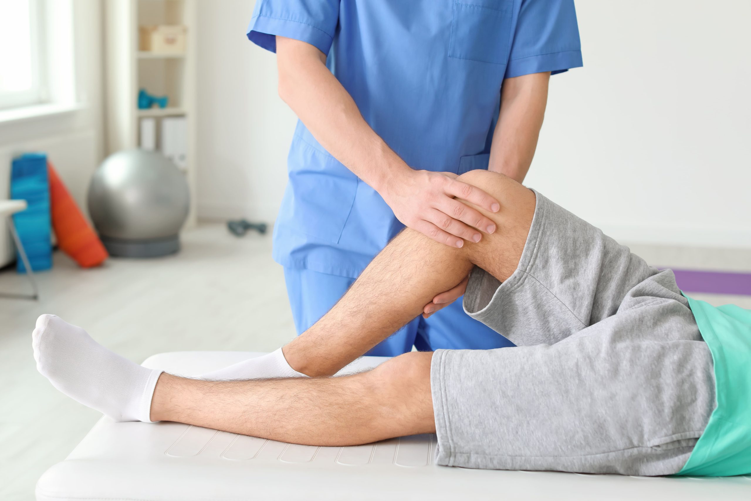
Many people searching for knee cartilage damage recovery time in Singapore want realistic timelines. Because knee cartilage does not heal in the same way as skin or muscle, recovery is rarely a matter of a few days.
Timelines vary widely depending on the nature of the injury and the chosen treatment pathway. While mild cartilage irritation may settle within six to twelve weeks of structured conservative rehabilitation, recovery following surgical repair or restoration typically ranges from three months for basic daily activities to nine to twelve months or longer for a safe return to high-impact sports. Understanding the factors that govern cartilage healing can help you navigate each stage of your recovery with confidence.
What Affects Knee Cartilage Damage Recovery Time?
No single timeline applies to everyone. Recovery depends on how large the damage is, where it sits in the knee, and which other structures are involved. Age, baseline fitness, and the chosen treatment path also play a significant part.
Timelines often follow recognisable patterns. Some people improve steadily over several weeks with guided physiotherapy and activity modification. Others require a structured, multi-month rehabilitation programme following cartilage surgery before they can safely return to running or competitive sport.
How Knee Cartilage Works and Why It Heals Slowly
Knee cartilage damage refers to a tear, thinning, or wearing away of the smooth, rubbery tissue (articular cartilage) covering the ends of the thigh bone (femur), shin bone (tibia), and kneecap (patella). It can result from an acute sports injury (such as a sudden pivot or collision) or develop gradually through repetitive wear. Common symptoms include pain deep in the joint, swelling, stiffness, and catching sensations.
Think of cartilage as the shock absorber in a car: it cushions joint loading during walking, jumping, and pivoting, allowing bones to glide smoothly over one another with minimal friction.
Most body tissues heal because circulating blood delivers oxygen, nutrients, and repair cells directly to the site of an injury. Articular cartilage, however, is avascular, meaning it has no direct blood supply. Instead, it relies on the slow diffusion of nutrients from the surrounding synovial fluid inside the joint. Because of this limited nutritional supply, minor surface damage heals slowly, and larger or deeper defects often cannot repair themselves fully without medical intervention.
Types of Cartilage Damage
Orthopaedic specialists generally grade cartilage damage according to depth:
- Mild: The cartilage surface becomes softened or swollen, though the structural surface remains intact.
- Moderate: Partial-thickness fissures or tears extend into the cartilage layer without reaching the underlying bone.
- Severe: Full-thickness cartilage loss occurs, exposing the underlying subchondral bone.
Mild to moderate damage frequently responds to non-surgical care over several weeks to a few months. Full-thickness focal defects typically take longer to resolve and may warrant surgical restoration.
Focal Defects vs Widespread Wear
A focal defect is an isolated, well-defined patch of cartilage damage surrounded by healthy tissue, comparable to a pothole in an otherwise smooth road. It often follows a specific traumatic injury, such as an ACL tear or a hard pivot on the football pitch. Orthopaedic surgeons can often repair, stimulate, or graft tissue in this type of isolated lesion.
Widespread wear, such as osteoarthritis, resembles a carpet thinning across an entire room. In these cases, recovery focuses on reducing inflammation, strengthening surrounding musculature, and protecting remaining joint surfaces, rather than surgically repairing a single localised spot.
Key Factors That Influence Knee Cartilage Injury Recovery
Evaluating these variables helps determine whether a patient is best suited to non-surgical rehabilitation or surgical intervention:
| Factor | How It Affects Healing | Clinical Management Approach |
|---|---|---|
| Defect Size | Larger defects carry higher mechanical loads and may take longer to heal or require surgical restoration | Accurate diagnostic imaging (MRI) guides treatment planning |
| Location | Weight-bearing zones on the femoral condyle tolerate load later than non-weight-bearing surfaces | Weight-bearing restrictions are tailored to the exact injury site |
| Age and Joint Health | Younger patients often possess greater cellular repair capacity; older joints may show broader degenerative change | Rehabilitation goals are matched to baseline joint condition |
| Activity Demands | High-impact, pivoting sports require higher tissue durability and longer rehabilitation than daily walking | Staged, milestone-based return-to-sport progression |
| Body Weight | Excess body weight increases compressive force across healing cartilage with every step | Gradual, sustainable load management and cross-training |
| Concomitant Injuries | Concurrent ligament (ACL) or meniscal tears can lengthen overall rehabilitation timelines | Addressing all structural injuries within one coordinated surgical or physical plan |
| Rehabilitation Adherence | Skipping exercises or progressing impact prematurely can compromise repair tissue | Consistent physiotherapy guided by clinical criteria |
Size and Location of the Cartilage Defect
Small defects often settle sooner because surrounding healthy tissue can help support joint loads. Larger defects place greater mechanical stress on surrounding cartilage. Location is equally important: a lesion on the central weight-bearing surface of the femur experiences continuous compressive force during walking, necessitating a longer period of protected weight-bearing than a lesion behind the kneecap.
Age, Activity Level, and Overall Joint Health
Younger patients generally have higher cellular activity that supports cartilage repair, though individual healing rates vary. A physically conditioned individual often recovers neuromuscular control faster than a deconditioned person. Similarly, the end-stage recovery goals of a recreational walker differ significantly from those of a competitive footballer.
Whether Other Knee Structures Are Also Injured
Cartilage injuries frequently occur alongside meniscus tears or ligament ruptures. Managing one problem while leaving another unaddressed can compromise joint mechanics and delay recovery. A comprehensive clinical assessment ensures that all damaged structures are rehabilitated in harmony.
Non-Surgical Recovery
Many individuals with partial-thickness cartilage damage or early degenerative wear experience meaningful improvement without surgery. This conservative approach typically involves:
- Relative rest and temporary offloading
- Targeted activity modification
- Structured physiotherapy
- Supportive bracing where appropriate
- Therapeutic joint injections in selected cases
Improvement is typically gradual, unfolding over several weeks to a few months.
Activity Modification and Load Management in the Early Weeks
During the initial one to two weeks, the primary goal is reducing pain and joint effusion (swelling). Activities that place high shear or compressive forces across the joint are temporarily modified, including:
- Running and jumping
- Deep unassisted squats and lunges
- Sudden cutting, pivoting, and twisting movements
This does not require complete bed rest. Low-impact movements such as gentle walking, swimming, or low-resistance stationary cycling help keep the leg muscles engaged and circulate synovial fluid without overloading healing cartilage.
Physiotherapy Milestones and Typical Timelines
A structured physiotherapy programme usually spans six to twelve weeks:
- Weeks 1 to 3: Focus on reducing joint swelling, restoring full knee extension and gentle flexion, and activating the quadriceps.
- Weeks 3 to 8: Focus on progressive strengthening of the quadriceps, hamstrings, and gluteal muscles, improving single-leg balance, and restoring a normal gait.
- Weeks 8 to 12 and Beyond: Gradual reintroduction of low-impact cardiovascular training, followed by sport-specific functional drills.
Strong supporting muscles act as biological shock absorbers, distributing joint loads away from damaged cartilage.
When Injections May Be Considered
If joint pain and stiffness persist despite rehabilitation, your doctor may discuss injection therapies:
- Hyaluronic acid injections: Aimed at supplementing joint fluid viscosity, improving lubrication, and easing mechanical friction.
- Platelet-rich plasma (PRP): Delivers a concentration of your own platelets and growth factors, which is intended to help soothe joint irritation and support symptom management.
Responses vary by individual, and suitability depends on clinical assessment.
Knee Cartilage Surgery Recovery Time
When conservative management does not adequately relieve symptoms, or when dealing with a full-thickness focal defect, surgical intervention may be considered. The table below outlines general recovery timelines across common procedures:
| Surgery Type | What It Involves | Recovery to Daily Activities | Recovery to Sport or High-Impact Activity | Key Clinical Considerations |
|---|---|---|---|---|
| Arthroscopic Debridement | Keyhole trimming and smoothing of loose or frayed cartilage flaps | 1 to 2 weeks | 6 to 12 weeks | Eases mechanical catching; does not regrow lost cartilage |
| Microfracture | Creating tiny perforations in the bone marrow to stimulate fibrocartilage repair | 6 to 8 weeks (with crutches) | 9 to 12 months | Best suited for smaller focal defects; repair tissue is fibrocartilage |
| Osteochondral Grafting (OATS) | Transferring cylinders of bone and healthy cartilage into the defect | 6 to 12 weeks | 6 to 12 months | Provides durable hyaline cartilage; requires graft incorporation |
Arthroscopic Debridement (Chondroplasty)
Arthroscopy is minimally invasive keyhole joint surgery performed through small skin incisions. In a debridement procedure, the surgeon trims unstable, frayed cartilage edges that cause catching or mechanical irritation. Recovery is typically fast: patients can often bear weight within days and return to light everyday activities within one to two weeks. Debridement relieves symptoms but does not replace worn cartilage.
Microfracture and Marrow Stimulation
Microfracture involves making tiny holes in the subchondral bone plate at the base of a cartilage defect. This allows blood and stem cells from the bone marrow to form a clot that matures into fibrocartilage repair tissue. Because this immature clot is fragile, protected weight-bearing on crutches is required for six to eight weeks. Returning to high-impact sports takes nine to twelve months as the repair tissue gradually consolidates.
Cartilage Grafting and Cell-Based Restoration (OATS)
Osteochondral autograft transfer (OATS) moves small plugs of healthy bone and cartilage from a non-weight-bearing part of the knee to fill the defect.
What a Typical Rehabilitation Timeline Looks Like
Rehabilitation is an active, milestone-driven process:
Weeks 1 to 6: Protecting the Repair and Restoring Range of Motion
The focus is on managing swelling, protecting healing repair tissue, and gently regaining joint flexion and extension. Depending on the surgical procedure and defect location, patients may use crutches and a knee brace to limit weight-bearing. Early passive range-of-motion exercises and isometric quadriceps activations begin promptly under professional guidance.
Weeks 6 to 12: Progressive Loading and Strength Building
Weight-bearing is gradually increased towards full capacity. Exercises progress to closed-chain movements, including leg presses, stationary cycling, pool exercise, and balance training. Rehabilitation targets the quadriceps, hamstrings, and hip abductors to stabilise the joint during walking.
Three Months Onward: Functional Conditioning and Sports Preparation
From three months, rehabilitation incorporates light jogging drills (once strength thresholds are met), agility training, and sport-specific movements. High-impact running and competitive sports clearance typically occurs between nine and twelve months, determined by functional testing and muscular symmetry rather than calendar time alone.
Signs That Suggest Recovery Is Progressing Well
- Joint swelling decreases progressively over weeks rather than fluctuating widely
- Knee flexion and extension improve steadily towards normal limits
- Walking pattern becomes smooth and symmetric without a limp
- Quadriceps and hamstring strength tests improve consistently at each clinical review
- Mild post-exercise soreness resolves within 24 hours
Your orthopaedic team will confirm whether functional milestones are being achieved safely.
How to Support Your Knee Cartilage Recovery at Every Stage
- Consistent physiotherapy attendance: Regularly completing prescribed exercises rebuilds joint stability and offloads healing tissue.
- Guided, low-impact movement: Gentle movement approved by your doctor promotes synovial fluid circulation and joint nourishment.
- Weight management: Maintaining a healthy body weight reduces cumulative mechanical load across the knee joint.
- Balanced nutrition and sleep: Adequate protein intake, balanced hydration, and quality sleep support the body’s natural tissue repair processes.
- Clear clinical communication: Informing your care team of sudden swelling, catching, or pain helps keep your recovery plan on track.
When to See a Knee Specialist
While minor aches often settle with rest, an orthopaedic evaluation from a knee specialist in Singapore is recommended if you experience:
- Knee swelling that persists beyond 48 hours
- Mechanical locking or catching that interrupts movement
- A sensation that the knee is unstable or giving way
- Inability to bear weight comfortably on the affected leg
- Joint pain that worsens despite rest and activity modification
An assessment provides clarity on the underlying structural cause, allowing you and your doctor to determine whether conservative management or surgical restoration is most appropriate.
Commonly Asked Questions
How long does it take to recover from knee cartilage damage without surgery?
Many individuals notice meaningful improvement within six to twelve weeks of structured physiotherapy, load management, and activity modification. Achieving full functional recovery for partial-thickness cartilage damage may take a few months, depending on defect size, baseline conditioning, and rehabilitation consistency.
Can damaged knee cartilage heal on its own?
Articular cartilage has very limited natural healing capacity because it lacks a direct blood supply. While minor surface softening may stabilise with rest, larger full-thickness defects rarely heal on their own. However, surrounding muscle strengthening and non-surgical care can often successfully resolve pain and restore function even if the cartilage does not fully regrow.
How soon can I return to sport after knee cartilage surgery?
Return-to-sport timelines depend heavily on the specific procedure. Following arthroscopic debridement, light sport may resume within six to twelve weeks. Following microfracture or cartilage grafting, returning to high-impact sport typically requires nine to twelve months or longer. Clearance is based on objective strength, balance, and functional testing.
Is knee cartilage damage permanent?
Because articular cartilage does not regenerate easily, some structural changes may remain. However, permanent structural changes do not mean permanent disability. With proper rehabilitation, targeted strengthening, and thoughtful load management, many active adults return to active sports and daily routines.
What happens if knee cartilage damage is left unmanaged?
While small, stable lesions may remain quiet for some time, unmanaged cartilage damage can gradually enlarge due to ongoing friction. Over time, progressive wear increases the risk of early secondary osteoarthritis. Early clinical assessment helps identify strategies to protect joint longevity.
Next Steps for Your Knee Cartilage Recovery
Knee cartilage damage recovery time depends on individual factors: defect size, anatomical location, patient age, baseline fitness, and whether concurrent ligament or meniscus injuries are present. An accurate clinical diagnosis and detailed imaging provide the foundation for a personalised recovery plan tailored to your lifestyle and athletic goals.
Disclaimer: This article is for informational purposes only and does not constitute medical advice. Please consult a qualified healthcare professional for personalised guidance.
Speak with a Knee Specialist About Your Recovery Plan
Consulting an orthopaedic specialist experienced in joint preservation and sports surgery allows you to explore the full spectrum of evidence-based options, from guided conservative rehabilitation to advanced cartilage restoration procedures.