
Call Us +65 6884 6788

September 2026

Twisting your ankle is one of those moments where everything stops. The pain hits, the swelling starts, and suddenly you are left wondering: is this serious, or will it sort itself out? Knowing which type of injury you are dealing with can make a real difference to how well and how quickly you recover.

Sprained Ankle or Broken Ankle: Why Getting the Right Answer Matters

Ankle injuries are among the most common musculoskeletal problems seen in Singapore, whether you are a weekend runner, a regular badminton player, or someone who simply stepped off a kerb awkwardly. A bad sprain and a fracture can look and feel remarkably similar in the first few hours after injury: both cause pain, swelling, and difficulty walking.

A missed fracture that goes untreated can lead to poor bone healing, long-term instability, or arthritis in the joint. An incorrectly managed sprain can result in chronic weakness and repeated re-injury.

What Happens Inside Your Ankle During an Injury

An ankle sprain occurs when the ligaments connecting the bones of the ankle are stretched or torn. An ankle fracture occurs when one or more of the bones forming the ankle joint crack or break completely. Both can happen during the same injury event, which is part of what makes self-diagnosis unreliable.

Your ankle is a complex joint made up of three bones: the tibia (shin bone), the fibula (the thinner bone running alongside it), and the talus (the bone sitting beneath them). These bones are held together and stabilised by ligaments, which you can think of as strong rubber bands connecting bone to bone. Surrounding muscles and tendons provide additional support and movement.

Ligament injuries (sprains)

When your ankle rolls inward or outward beyond its normal range of motion, the ligaments on the outer or inner side of the ankle are forced to stretch further than they are designed to. Depending on how far they are pushed, the fibres may stretch slightly, partially tear, or rupture completely. The most commonly affected ligament in an ankle sprain is the anterior talofibular ligament (ATFL), located on the outer side of the ankle. Ligament injuries do not show up on X-ray, which is one reason imaging alone does not always give the full picture.

Bone injuries (fractures)

A fracture occurs when the force applied to the ankle exceeds what the bone can absorb. This can happen during a high-impact fall, a direct blow, or even a severe twisting motion. The fibula is the most frequently fractured bone in ankle injuries. In some cases, particularly with repetitive loading rather than a single event, stress fractures can develop gradually without any obvious trauma. These hairline cracks are easy to miss but can worsen significantly if activity continues without treatment.

How to Tell the Difference Between a Sprained Ankle and a Broken Ankle

There is considerable overlap between the two injuries. While certain signs can guide your initial assessment, only a clinical examination combined with imaging can confirm which injury you have sustained.

The table below compares the key features of each injury type as a starting point for your own assessment.

Feature Sprained Ankle Broken Ankle
Pain location Along the outer or inner ankle, over the ligament Directly over the bone, often the fibula or medial malleolus
Swelling pattern Soft tissue swelling around the joint Can be more focal or involve visible deformity
Ability to bear weight Often possible, though painful Frequently impossible, especially with displaced fractures
Bruising Common, may appear within hours Common, often more extensive
Deformity Rarely visible Possible if bones are displaced
Sound at time of injury May hear a pop or snap from the ligament May hear a crack or crunch

Pain and tenderness patterns

With a sprain, tenderness is typically felt over the soft tissue on the outer or inner side of the ankle, directly over the affected ligament. With a fracture, tenderness tends to be concentrated directly over the bone itself, particularly along the fibula or at the bony prominences on either side of the ankle (the medial and lateral malleoli). If pressing on a specific bony point produces sharp, localised pain, that is a signal worth discussing with a healthcare professional.

Swelling, bruising, and visible changes

Both injuries cause swelling and can produce significant bruising. Bruising from a sprain typically appears around the soft tissue of the outer ankle. With a fracture, bruising may be more widespread and can track down towards the foot. Visible deformity, where the ankle appears out of its normal alignment, may indicate a fracture and requires prompt medical attention. Do not attempt to push or manipulate the ankle back into position.

Can you put weight on it?

The ability to bear weight is one of the most commonly used early indicators, but it is not a reliable rule on its own. Some people with significant fractures manage to limp a short distance, while others with severe ligament tears cannot put any weight on the foot at all. The Ottawa Ankle Rules, a clinical decision guide used by doctors (explained further below), incorporate weight-bearing ability as one factor among several . If bearing weight causes sharp, intense pain or feels mechanically unstable, seeking assessment promptly is advisable rather than testing it further.

Ankle Injury Severity: Grading Sprains and Classifying Fractures

Not all sprains are minor, and not all fractures require surgery. The severity of your injury determines whether conservative care at home is sufficient or whether specialist intervention is needed.

Sprain Grade Description Typical Symptoms General Recovery
Grade I (Mild) Minor stretching of ligament fibres, no tearing Mild pain, slight swelling, minimal functional loss 1 to 2 weeks
Grade II (Moderate) Partial tear of the ligament Moderate pain, noticeable swelling and bruising, some instability 3 to 6 weeks
Grade III (Severe) Complete rupture of the ligament Significant pain, marked swelling, joint instability, difficulty bearing weight 6 to 12 weeks or longer

Fractures are classified in several ways. A stable fracture means the bone has cracked but the fragments remain in their correct position. A displaced fracture means the bone ends have shifted out of alignment. Stress fractures are hairline cracks that develop gradually from repetitive loading rather than a single traumatic event. Each type carries different implications for treatment and the time needed to heal.

Common Causes of Ankle Sprains and Fractures in Singapore

Sports and exercise injuries

Sports involving rapid changes of direction, jumping, or uneven terrain carry a higher ankle injury risk. Football, basketball, badminton, and running are among the more frequent causes seen in orthopaedic clinics here. Trail runners at MacRitchie Reservoir or Bukit Timah Nature Reserve face additional risk from roots, rocks, and unpredictable ground surfaces. Even lower-intensity activities like hiking or recreational cycling can result in a rolled ankle if the terrain is uneven or if footwear is inadequate.

Everyday slips and falls

Not every ankle injury happens during sport. Wet hawker centre floors, uneven pavement edges, poorly lit stairwells, and stepping off escalators at an angle are all everyday hazards in Singapore. Wearing unsupportive footwear, such as flat sandals or worn-out shoes, increases the risk further.

What to Do Immediately After an Ankle Injury

The actions you take in the first 48 to 72 hours after an ankle injury can meaningfully influence how well your ankle heals. The PRICE protocol provides a straightforward framework.

First-aid steps you can take at home

  1. P: Protection. Avoid putting full weight on the injured ankle. Use crutches or support from another person if you need to move.
  2. R: Rest. Reduce activity to allow the initial inflammatory response to settle. This does not mean complete bed rest, but it does mean avoiding the activity that caused the injury.
  3. I: Ice. Apply an ice pack wrapped in a cloth to the injured area for 15 to 20 minutes at a time, every two to three hours. Never apply ice directly to skin.
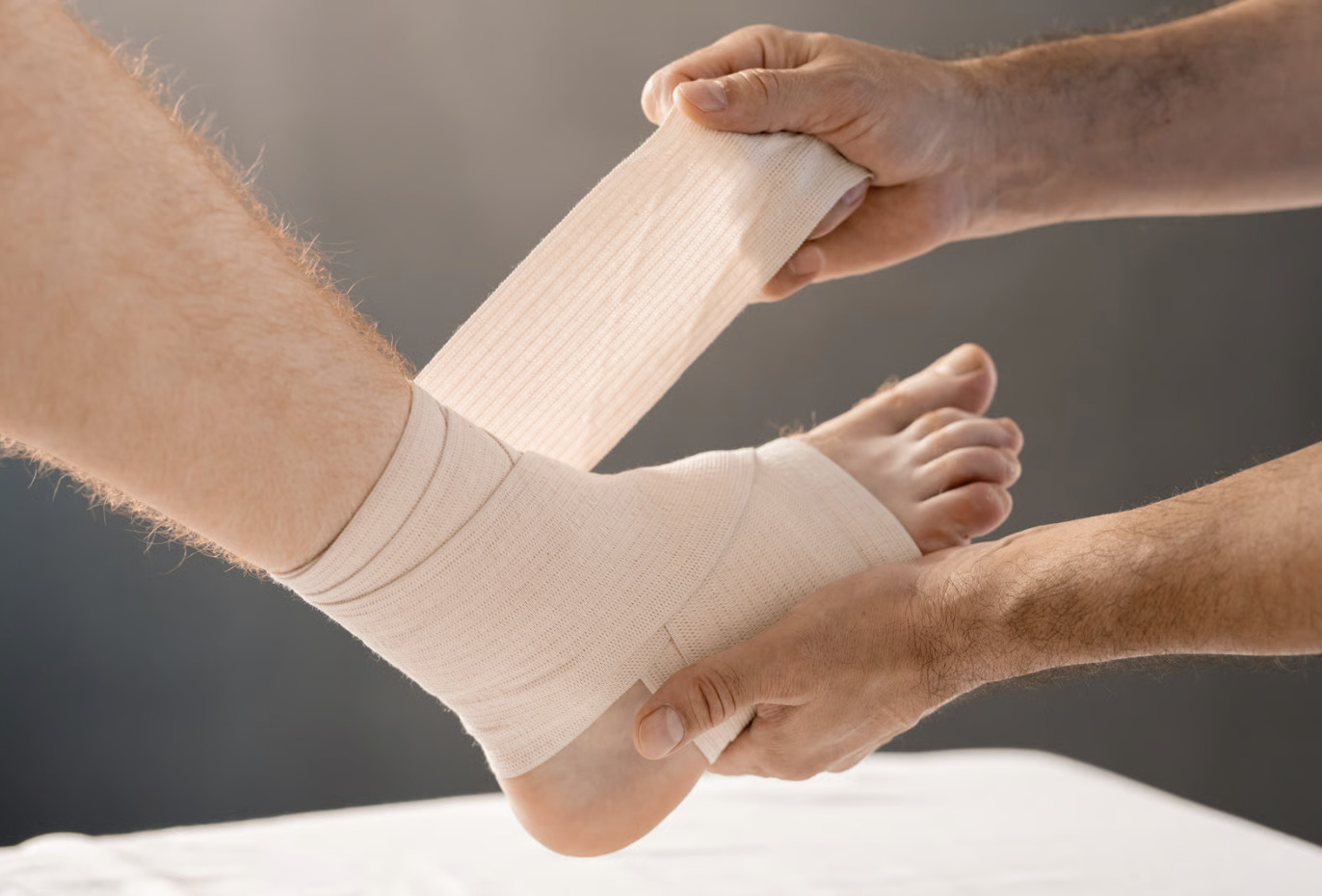
  4. C: Compression. A compression bandage applied around the ankle helps limit swelling. Wrap firmly but not so tightly that it restricts circulation.
  5. E: Elevation. Raise your ankle above the level of your heart when resting, using a pillow or cushion. This helps reduce swelling by encouraging fluid to drain away from the injury site.

What to avoid in the first 48 hours

  • Applying heat to the ankle (hot water bottles, heat patches, or hot showers directed at the injury)
  • Drinking alcohol, which can increase swelling
  • Massaging the injured area vigorously
  • Returning to sport or exercise before the ankle has been assessed
  • Walking through significant pain without support

Quick first-aid checklist for ankle injuries

  • Stop activity immediately
  • Protect the ankle from further stress
  • Apply ice (wrapped in cloth) for 15 to 20 minutes every 2 to 3 hours
  • Apply a compression bandage
  • Elevate the ankle above heart level
  • Avoid heat, massage, and alcohol
  • Seek medical assessment if pain is severe, deformity is visible, or weight-bearing is impossible

How Ankle Injuries Are Diagnosed in Singapore

When you attend a clinic for an ankle injury, the assessment typically begins with a physical examination. Your doctor will ask how the injury happened, where the pain is most intense, and whether you were able to bear weight immediately after the incident. They will then gently palpate (press on) specific points around the ankle to identify areas of tenderness.

Many doctors use the Ottawa Ankle Rules as a clinical decision guide to determine whether an X-ray is necessary. These rules identify specific bony landmarks around the ankle and foot: if pressing on those points causes pain, or if you cannot take four steps immediately after the injury, an X-ray is recommended. This helps avoid unnecessary imaging for minor soft tissue injuries while ensuring fractures are not missed.

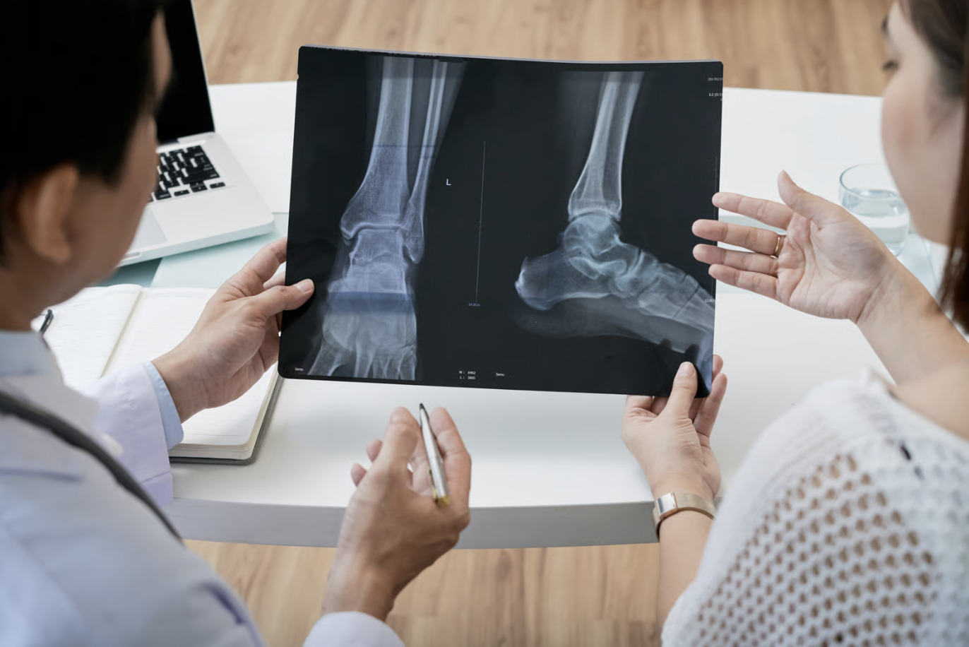
An X-ray is the standard first imaging step and will clearly show most fractures. However, ligament injuries, cartilage damage, and stress fractures in their early stages do not always appear on X-ray. In these cases, your doctor may recommend an MRI (magnetic resonance imaging) scan, which produces detailed images of soft tissue structures and can identify injuries that plain X-rays may miss.

How Sprained Ankles and Broken Ankles Are Treated

Treatment for ankle injuries ranges from home-based management to surgical intervention, depending on the type and severity of the injury.

Conservative treatment for sprains

Most ankle sprains, including most Grade I and Grade II injuries, may respond well to conservative management. This typically involves a structured physiotherapy programme to restore range of motion, strength, and proprioception (balance and coordination). A supportive ankle brace may be recommended during the early recovery phase and when returning to sport. For moderate sprains, a short period in a walking boot can help reduce pain while allowing gradual weight-bearing.

Anti-inflammatory medication may be suggested to manage pain and swelling in the acute phase; this should be discussed with your doctor. For Grade III sprains, many patients may achieve good outcomes with physiotherapy and bracing rather than surgery, though this depends on individual circumstances.

When a fracture needs surgery

Stable fractures, where the bone fragments remain correctly positioned, are often managed with immobilisation in a cast or walking boot and a period of protected weight-bearing. Displaced fractures, where the bone ends have shifted out of alignment, may require surgical fixation to restore the correct anatomy and allow the joint to heal properly. Surgery may involve the placement of plates and screws to hold the bone fragments in position while healing occurs.

For certain ankle conditions involving ligament instability or cartilage damage alongside a fracture, arthroscopic techniques (keyhole surgery using a small camera and instruments inserted through tiny incisions) may be used. These approaches allow the surgeon to address damage inside the joint with minimal disruption to surrounding tissue.

Rehabilitation and returning to activity

Regardless of whether your ankle injury is managed conservatively or surgically, rehabilitation is an important part of long-term recovery. A graded physiotherapy programme addresses not just pain and swelling but the underlying strength deficits and balance impairments that, if left unaddressed, may increase the risk of re-injury. Returning to sport before rehabilitation is complete is one of the more common reasons patients experience recurrent ankle sprains.

Recovery Timeline: What to Expect After a Sprained or Broken Ankle

Healing timelines vary considerably from person to person. Factors such as age, baseline fitness, the presence of other health conditions, and how closely a rehabilitation programme is followed all influence how quickly recovery progresses.

Injury Type Expected Recovery Range
Mild sprain (Grade I) 1 to 2 weeks
Moderate sprain (Grade II) 3 to 6 weeks
Severe sprain (Grade III) 6 to 12 weeks, sometimes longer
Stable fracture (non-surgical) 6 to 10 weeks
Fracture requiring surgery 3 to 6 months (including rehabilitation)

Pain going away does not always mean the ankle is fully healed. Ligaments and bone require time to remodel and regain their full strength, and the balance and coordination systems of the ankle benefit from deliberate retraining. Stopping rehabilitation as soon as the pain resolves is one of the more common reasons patients experience setbacks.

Protecting Your Ankles and Reducing Re-Injury Risk

A previous ankle sprain is associated with an increased likelihood of future ankle sprains. Targeted prevention strategies may help reduce that risk.

  • Strengthening exercises: Calf raises, resistance band exercises, and single-leg strengthening can help build the muscular support around the ankle joint.
  • Proprioception training: Balance and coordination retraining, such as single-leg standing, wobble board exercises, and dynamic balance drills, retrains the nerve pathways that help your ankle respond to unstable surfaces.
  • Appropriate footwear: Wear supportive shoes suited to your activity. Avoid worn-out trainers for running or sport, and choose footwear with adequate ankle support for trail activities.
  • Warm up before exercise: A proper warm-up that includes dynamic movements helps prepare the ankle ligaments and surrounding muscles for the demands of sport.
  • Ankle bracing during return to sport: For those with a history of sprains, wearing a supportive ankle brace during higher-risk activities may provide an additional layer of protection while strength and proprioception are still being rebuilt.

When to Seek Professional Help

  • You are unable to bear weight on the ankle at all
  • There is visible deformity or the ankle appears out of alignment
  • Swelling is severe and does not begin to improve after 48 to 72 hours
  • Pain remains significant beyond a few days despite rest and first aid
  • You have experienced repeated ankle sprains on the same side
  • You heard a distinct crack or crunch at the time of injury
  • You are an athlete who needs to return to training or competition safely

Commonly Asked Questions

Is it possible to walk on a broken ankle without realising it?

Yes, this can occur. Certain stable fractures, particularly those involving the fibula without displacement, may allow limited weight-bearing with pain that feels manageable. Stress fractures in particular often develop gradually with only mild discomfort at first. Persistent ankle pain after an injury should be assessed by a healthcare professional rather than assumed to be a minor sprain.

How long should I wait before seeing a doctor for a swollen ankle?

If swelling is severe, you cannot bear weight, or there is any visible deformity, seeking assessment promptly is advisable rather than waiting. For milder swelling with manageable pain, applying the PRICE protocol for 24 to 48 hours is reasonable. If swelling is not improving or pain is worsening after that period, a medical review is advisable.

Can a severe sprain be worse than a fracture?

In terms of recovery time and long-term impact, this is possible. A Grade III complete ligament rupture can take longer to rehabilitate than a simple stable fracture, and if not treated properly, can lead to chronic ankle instability that affects daily activities and sport. Fractures, once healed, may restore normal joint mechanics more predictably than severely torn ligaments that have not been adequately rehabilitated, though outcomes vary by individual.

Do I need an MRI for an ankle injury, or is an X-ray enough?

An X-ray is the appropriate first step for most ankle injuries, as it can effectively identify fractures. If the X-ray is clear but pain and swelling persist, or if your doctor suspects ligament damage, cartilage injury, or a stress fracture not visible on plain film, an MRI provides more detailed information about the soft tissue structures inside the joint. A healthcare professional can advise whether further imaging is needed based on your clinical picture.

How can I prevent ankle sprains when playing sports in Singapore?

Wearing sport-specific footwear with adequate ankle support, and replacing trainers when the sole shows significant wear, may help reduce injury risk. Incorporating proprioception and balance training into your regular exercise routine is also worth considering. Warming up thoroughly before matches and training sessions is advisable. On outdoor surfaces such as trail paths at MacRitchie or Bukit Timah, reducing your pace on uneven or wet terrain and staying attentive to the ground ahead can help. For those with a history of sprains, speaking with a healthcare professional about ankle bracing options may be helpful.

Next Steps

Whether you have just twisted your ankle or are dealing with a recurring issue that never quite resolved, applying first aid promptly, monitoring your symptoms, and seeking assessment when the signs suggest something more than a minor sprain are all reasonable steps. Early, accurate diagnosis of ankle injuries supports better outcomes and a more complete return to the activities you enjoy.

Speak with an Orthopaedic Specialist About Your Ankle Injury

If you are unsure about your ankle injury or would like a thorough assessment from a specialist with experience in sports and musculoskeletal conditions in Singapore, you may wish to arrange a consultation to discuss your symptoms, review any imaging, and explore your treatment and rehabilitation options.

Disclaimer: This article is for informational purposes only and does not constitute medical advice. Please consult a qualified healthcare professional for personalised guidance.

Book Appointment

Fill up the form and we will get back to you soon!

    Synergy Orthopaedic Group

    September 2026

    That sharp, stabbing pain in your heel the moment you take your first step out of bed catches many people off guard. Plantar fasciitis is one of the most common foot complaints seen in orthopaedic practice in Singapore, affecting everyone from office workers to recreational runners.

    Understanding Morning Heel Pain and Plantar Fasciitis

    You wake up, swing your legs over the side of the bed, and the moment your foot touches the floor, a sharp pain shoots through your heel. It can ease after a few minutes of walking, only to return after sitting for a while.

    This pattern is a hallmark of plantar fasciitis. Many patients recover with the right approach.

    What Is Plantar Fasciitis?

    Plantar fasciitis is inflammation of the plantar fascia, the thick band of tissue that runs along the sole of your foot, connecting your heel bone to the base of your toes. “Fasciitis” simply means inflammation of the fascia.

    When this band is placed under repetitive stress, small micro-tears can develop in the tissue. These micro-tears trigger an inflammatory response (your body’s natural repair mechanism), causing pain and stiffness, particularly at the point where the fascia attaches to the heel bone.

    Think of the plantar fascia like a bowstring stretched along the underside of your foot. Every step you take loads that string. If the load exceeds what the tissue can handle, whether through a sudden increase in activity, prolonged standing, or structural factors, the fibres begin to fray at their weakest point.

    Plantar fasciitis is common among runners and sports enthusiasts, but it is equally seen in those who spend long hours on their feet at work, or those who have recently increased their activity levels after a sedentary period. It is a condition that crosses all ages and fitness levels.

    Why Does Plantar Fasciitis Cause Heel Pain in the Morning?

    When you are at rest, the plantar fascia naturally shortens and contracts. The longer you rest, the tighter the tissue becomes.

    The moment you stand up and place weight on your foot, that contracted tissue is suddenly stretched and loaded all at once. This abrupt tension on already-irritated fibres produces that sharp, first-step pain.

    As you walk around and the tissue warms up and loosens, the pain often eases. Many people describe the pain as worst first thing in the morning or after sitting for an extended period, then improving after a few minutes of movement. After prolonged activity, the pain can return as the tissue fatigues again.

    This cycle of pain, relief with movement, and return after rest is one of the clearer indicators that plantar fasciitis may be the cause of your heel discomfort.

    Common Causes and Risk Factors

    Plantar fasciitis typically develops when a combination of factors places more load on the plantar fascia than it can comfortably handle.

    Lifestyle and Occupational Factors

    • Prolonged standing or walking: Jobs that require long hours on hard floors, such as healthcare, retail, or food service, place continuous load on the tissue.
    • Flat, unsupportive footwear: In Singapore’s warm climate, slippers and flat sandals are popular choices, but they offer minimal arch support and cushioning, which can increase stress on the plantar fascia over time.
    • Sudden weight changes: An increase in body weight raises the load on the foot’s soft tissues with every step.
    • Hard flooring surfaces: Tile and concrete floors, common in Singapore homes and workplaces, offer little shock absorption compared to softer surfaces.

    Foot Structure and Biomechanics

    • Flat feet (low arches): When the arch collapses with each step, the plantar fascia is stretched further than it is designed to handle.
    • High arches: A rigid, high-arched foot distributes load unevenly, concentrating stress on the heel and ball of the foot.
    • Tight calf muscles and Achilles tendon: When the calf muscles (the muscles at the back of your lower leg) are tight, they pull on the heel bone and increase tension in the plantar fascia.
    • Overpronation: This is the tendency of the foot to roll inward excessively with each step, placing uneven stress on the fascia.

    Sports and Training-Related Triggers

    • Running and high-impact sports: Repetitive loading from running, football, basketball, and similar activities can overload the plantar fascia, especially on hard surfaces.
    • Sudden increases in training volume: Ramping up your mileage or training intensity too quickly may not give the tissue adequate time to adapt.
    • Inadequate warm-up or cool-down: Skipping stretching before and after exercise keeps the calf muscles and plantar fascia tight and may increase vulnerability to injury.
    • Worn-out training shoes: Shoes that have lost their cushioning and support may no longer protect the foot adequately.

    How Is Plantar Fasciitis Diagnosed in Singapore?

    Diagnosis is usually straightforward and does not require complex investigations. An orthopaedic specialist will begin with a detailed clinical history, asking about the nature and timing of your pain, your activity levels, footwear habits, and how long symptoms have been present.

    A physical examination follows, with the doctor assessing tenderness at the medial calcaneal tubercle (the bony point on the inner side of your heel where the plantar fascia attaches). Reproducing your pain with gentle pressure at this specific point is a useful diagnostic indicator.

    Imaging may be recommended in certain situations:

    • Ultrasound: This can confirm thickening or tearing of the plantar fascia and is useful when the diagnosis is uncertain or to guide injection treatments.
    • X-ray: Occasionally used to identify a heel spur, which is a bony outgrowth on the heel bone. Heel spurs visible on X-ray are not always the source of pain. Many people have heel spurs without any symptoms. The plantar fascia inflammation, rather than the spur itself, is often responsible for the discomfort.

    In many cases, a thorough clinical assessment is sufficient to support the diagnosis and begin treatment.

    Plantar Fasciitis Treatment Options

    Treatment for plantar fasciitis is stepwise, starting with the least invasive approaches and progressing only if symptoms do not improve adequately. Many cases respond to conservative, non-surgical care.

    The table below summarises the main treatment approaches, what each involves, the typical timeline for improvement, and who each option may suit best.

    Treatment Type What It Involves Typical Timeline for Improvement Who It May Suit Best
    Rest and stretching Reducing aggravating activities; daily calf and plantar fascia stretches 4-8 weeks with consistent effort Most patients as a first-line approach
    Footwear modification Switching to supportive shoes; avoiding flat slippers and hard-soled footwear Improvement alongside other measures Anyone whose footwear may be contributing
    Physiotherapy Guided stretching, strengthening, manual therapy, and gait assessment 6-12 weeks Patients with biomechanical contributors or persistent symptoms
    Orthotics Custom or off-the-shelf insoles to support the arch and reduce fascial strain Improvement alongside other measures Those with flat feet, high arches, or overpronation
    Shockwave therapy Non-invasive sound wave treatment to stimulate tissue healing 3-6 sessions over several weeks Patients with symptoms lasting 3 months or more
    Corticosteroid injection Anti-inflammatory injection into the affected area Days to weeks for pain relief Short-term relief when pain is severe; used selectively
    Surgery Partial release of the plantar fascia; rarely required Several months of recovery Cases that have not responded to 6-12 months of conservative treatment

    Stretching, Rest, and Footwear Modifications

    The foundation of plantar fasciitis treatment is a consistent stretching routine combined with sensible footwear choices. Stretching the calf muscles and the plantar fascia may reduce tension on the heel attachment and support tissue healing.

    Key stretches include:

    • Plantar fascia stretch: While seated, cross one foot over the opposite knee and gently pull the toes back toward the shin. Hold for 20-30 seconds and repeat several times.
    • Calf stretch: Stand facing a wall with one foot forward and one foot back. Keep the back heel flat on the floor and lean into the wall to stretch the calf. Hold for 30 seconds on each side.

    Performing these stretches before taking your first steps in the morning may be helpful for many people. You may wish to discuss a stretching routine with your healthcare professional to confirm it is appropriate for your situation.

    Supportive shoes with good arch support and cushioned soles may help reduce the load on the plantar fascia. In Singapore’s climate, replacing flat slippers with sandals that offer arch support and cushioning can be a practical change worth considering.

    Physiotherapy and Orthotics

    A physiotherapist can assess the way you walk and move, identify contributing biomechanical factors, and guide you through a structured rehabilitation programme. This typically includes strengthening the foot and calf muscles, improving flexibility, and addressing any gait abnormalities.

    Custom or prefabricated orthotics (supportive insoles placed inside your shoes) may help correct foot alignment and reduce strain on the plantar fascia. They are often considered for those with flat feet, high arches, or overpronation.

    Shockwave Therapy and Injection Treatments

    For cases that persist beyond three months despite consistent conservative care shockwave therapy may be considered. This non-invasive treatment delivers targeted sound waves to the affected tissue, with the aim of stimulating the body’s healing response and reducing chronic inflammation.

    Corticosteroid injections (anti-inflammatory medication injected directly into the painful area) may provide short-term pain relief, particularly when symptoms are severe. They are used selectively, as repeated injections carry a small risk of weakening the plantar fascia over time.

    When Surgery May Be Considered

    Surgery for plantar fasciitis is uncommon. It is generally considered only when symptoms have persisted for six to twelve months despite thorough conservative treatment. The procedure typically involves a partial release of the plantar fascia to reduce tension at the heel attachment. Recovery takes several months, and the decision to proceed is made carefully on an individual basis.

    How Long Does Plantar Fasciitis Take to Heal?

    Many people with plantar fasciitis see meaningful improvement within six to twelve months with consistent conservative treatment. Some experience relief sooner, particularly when they begin stretching early and address contributing factors like footwear and activity levels. Recovery time varies by individual.

    Recovery time is influenced by several factors:

    • How long symptoms have been present: Earlier intervention may support faster recovery.
    • Consistency with stretching: Daily stretching may make a meaningful difference. Sporadic effort tends to produce slower results.
    • Activity modification: Continuing high-impact activities without modification can slow healing.
    • Individual factors: Foot structure, overall health, and adherence to the treatment plan all play a role.

    Progress is often measured in weeks and months, not days. Staying consistent with your rehabilitation, even on days when the pain feels manageable, is important for recovery.

    Three Daily Habits That May Support Recovery

    • Stretch before your first step: Performing calf and plantar fascia stretches while still in bed, before placing weight on your foot, may help reduce first-step pain.
    • Choose supportive footwear every day: Avoiding flat slippers around the house and wearing shoes or sandals with arch support and cushioning from the moment you get up may help reduce strain on the plantar fascia.
    • Gradually ease back into activity: Avoiding sudden spikes in walking, running, or standing duration and increasing activity levels incrementally may allow the tissue to adapt more comfortably.

    Returning to Exercise and Sports After Plantar Fasciitis

    Returning to running, gym training, or competitive sport after plantar fasciitis is a realistic goal for many people. A graduated return to activity means progressively increasing the load on the plantar fascia over time, giving the tissue a chance to adapt and strengthen. Returning to your previous training volume too quickly is one of the more common reasons for setbacks.

    During recovery, consider cross-training alternatives that maintain your fitness while reducing impact on the heel:

    • Swimming: Cardiovascular conditioning with no impact load on the foot.
    • Cycling: Low-impact and useful for maintaining leg strength and aerobic fitness.
    • Pool running: Allows you to maintain running mechanics without ground impact.

    Signs that you may be ready to return to higher-impact activities include:

    • No pain during normal daily walking
    • Minimal or no discomfort with gentle jogging on a soft surface
    • Consistent improvement in morning stiffness over several weeks

    If pain returns when you increase your activity, scaling back and allowing more recovery time before trying again may be helpful. Working with a physiotherapist to structure your return-to-sport plan may reduce the risk of re-injury.

    When to Seek Professional Help

    • Heel pain has persisted for more than two to four weeks despite rest, stretching, and footwear changes
    • Pain is worsening rather than gradually improving
    • You are unable to bear weight comfortably on the affected foot
    • Heel pain is significantly affecting your ability to walk, work, or participate in daily activities
    • You are an athlete and pain is preventing you from training or competing
    • You have tried conservative measures consistently but are not seeing progress

    Seeking evaluation early, rather than waiting for symptoms to become severe, may support simpler treatment and more straightforward recovery.

    Commonly Asked Questions

    Can plantar fasciitis go away on its own without treatment?
    Mild cases of plantar fasciitis can improve over time with rest alone, but recovery without any active management is typically slower and less predictable. Consistent stretching, footwear changes, and activity modification may improve outcomes. Ignoring persistent symptoms and continuing aggravating activities can allow the condition to become more chronic and harder to treat.

    Is plantar fasciitis more common in runners?
    Runners are at higher risk due to the repetitive impact loading involved, but plantar fasciitis is by no means exclusive to runners. It is common among people who stand for long hours, those who have recently increased their activity levels, and individuals with certain foot structures.

    Are heel spurs the same as plantar fasciitis?
    No. A heel spur is a bony outgrowth on the heel bone that may appear on an X-ray. Plantar fasciitis refers to inflammation of the plantar fascia tissue. The two can occur together, but many people have heel spurs without any pain, and many people with plantar fasciitis do not have heel spurs. The tissue inflammation, rather than the bony spur, is typically the source of the pain.

    Should I avoid walking if I have plantar fasciitis?
    Complete rest is generally not necessary and is not always recommended. Moderate walking is usually fine and may help maintain tissue mobility. Avoiding prolonged walking on hard surfaces, walking barefoot, or sudden increases in walking duration that overload the tissue may be helpful. Wearing supportive footwear and pacing your activity are generally considered more important than stopping movement entirely. You may wish to discuss activity modification with your healthcare professional.

    What type of shoes are best for plantar fasciitis in Singapore’s climate?
    Shoes or sandals with good arch support, a cushioned midsole, and a slight heel elevation (not completely flat) are generally worth considering. In Singapore’s warm climate, supportive sandals with contoured footbeds may be a practical option for everyday wear. Flat flip-flops, hard-soled shoes, and walking barefoot on hard floors, particularly first thing in the morning, are generally best avoided. A healthcare professional can provide guidance on footwear suited to your specific foot structure.

    Next Steps for Managing Your Heel Pain

    You may wish to begin with daily calf and plantar fascia stretches, review your footwear choices, and moderate any activities that are aggravating your symptoms.

    Monitor your progress over the next two to four weeks. Many people notice improvement with these foundational steps. If your pain is not responding, is worsening, or is limiting your daily function or training, arranging a specialist assessment may be a sensible next step.

    Disclaimer: This article is for informational purposes only and does not constitute medical advice. Please consult a qualified healthcare professional for personalised guidance.

    Speak with an Orthopaedic Specialist About Your Heel Pain

    If conservative measures have not provided adequate relief, or if you are unsure whether your heel pain is plantar fasciitis, a consultation with an orthopaedic specialist may help provide clarity. An accurate diagnosis and a structured plantar fasciitis treatment plan tailored to your needs are the foundation of effective recovery, supporting your return to daily activities and the things you enjoy.

    Book Appointment

    Fill up the form and we will get back to you soon!

      Synergy Orthopaedic Group

      back to top